
Our GFP/GMP Products · ANOVA Germany
MSC Secretome (MSEC) Germany
MSEC (mesenchymal stem cell secretome) is a cell-free preparation made from your own stem cells. It contains the molecules those cells release — exosomes, microvesicles, growth factors and cytokines — but no living cells. ANOVA IRM manufactures MSEC in Offenbach, Germany, under official authorisation and inspection by the German medicines authority. MSEC is experimental: there is no marketing authorisation and no phase 3 proof of efficacy for this product.
Because MSEC contains no cells, it can be frozen without losing its composition. One tissue collection therefore yields several doses that can be given repeatedly over an extended period — which is what chronic conditions require, and what living cells cannot easily provide. External laboratory and animal studies indicate that much of what mesenchymal stem cells (MSCs) do in the body is done by what they release rather than by the cells themselves. MSEC delivers those released factors without transferring cultured cells. Treatment is given as an individual therapeutic attempt (individueller Heilversuch) after a personal medical assessment. ANOVA IRM also offers Bone Marrow Concentrate (BMC); which product is appropriate is decided case by case. See the conditions we treat, how ANOVA IRM is regulated, and who we are.
| Component | Detail |
|---|---|
| Two products | Bone Marrow Concentrate (BMC) and Mesenchymal Stem Cell Secretome (MSEC), each manufactured under official authorisation; no marketing authorisation (experimental) |
| Authorisations | BMC § 20b / § 13 AMG · MSEC § 20b / § 20c AMG (German Medicines Act) |
| Location | Offenbach am Main, Germany |
What MSC, secretome and exosomes are, how a cell-free product compares with living cells, and the most common questions.
You are hereIntroduction: Stem Cells, Secretome and Exosomes – Need to Know
In this section:
- What are Mesenchymal Stem Cells (MSC)?
- Why are MSEC - Mesenchymal Stem Cells - More Often Used Compared Other Stem Cell Types
- What is a Stem Cell Secretome? What are Exosomes?
- What is a Stem Cell Secretome Therapy?
- Stem Cells or Secretome? Pros and Cons
- Where a Cell-Free Product Carries Less Risk
- Frequently Asked Questions — Secretome and Exosomes
What are Mesenchymal Stem Cells (MSC)?
Mesenchymal Stem Cells (MSCs) are one of the most widely studied stem cell types in medicine. They are being investigated in research for a wide range of diseases. The available clinical data are still early and limited, so neither efficacy nor safety in patients can be regarded as established.
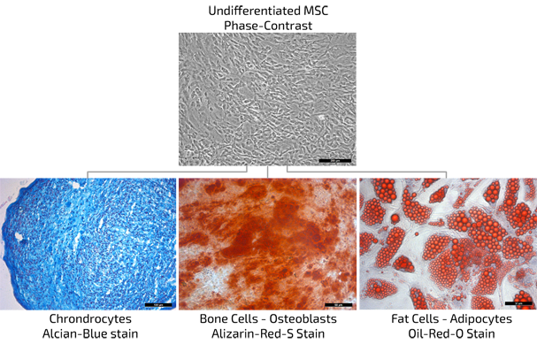
At the same time several hundred clinical studies are ongoing to investigate their possible regenerative effects in greater detail. From a biological point-of-view, they are progenitor cells of connective tissues. This means that they are important for building and maintaining the healthy status of connective tissues throughout the whole body, and are known as stromal stem cells. Likewise, they possess the ability to differentiate into a spectrum of other cell types, including chondrocytes (cartilage cells), osteoblasts (bone cells), adipocytes (fat cells), etc. This property classifies them as “multipotent stem cells” (being able to transform into many cell types).
Why are MSEC - Mesenchymal Stem Cells - More Often Used Compared Other Stem Cell Types
There are several reasons why MSC are among the most widely used stem cell types in medical applications and clinical trials in humans:
- They are relatively easy to harvest and be isolated from: Fat tissue, known as adMSCs (adipose derived MSCs) Bone Marrow, known as bmMSCs (bone marrow MSCs) Umbilical Cord, known as ucMSC (umbilical cord MSCs)
- MSCs are relatively stable to culture and expand in the laboratory
- They can go through relatively many division cycles without losing their expression profile, health and division potential
- Numerous animal, clinical and scientific data collected for various disease models, including neurodegenerative diseases
- Laboratory studies so far report a low tendency of MSCs to form tumour cells; long-term safety in patients is still being investigated
- Recent laboratory research is investigating the stem cell secretome profile, which has shown a range of regenerative and protective activities in preclinical models
- For these reasons, MSC are our preferred source for producing MSEC.
What is a Stem Cell Secretome? What are Exosomes?
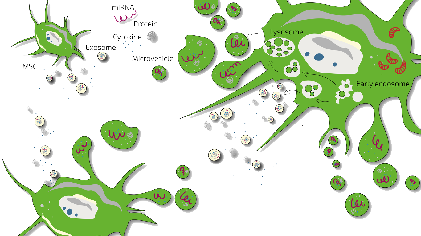
In scientific terms, the secretome is the totality of all substances released by stem cells to the outside. These include micro-vesicles, exosomes, proteins, cytokines (cell messengers), hormone-like substances and so-called immunomodulatory substances.
Based on current science, many researchers assume that the effect of stem cells in clinical trials to date does not come from the cells as such. It is thought to come from the vesicles, protein molecules and other substances such as cytokines that the cells release. These substances released to the outside are called "the secretome" as an umbrella term, because the release is called secretion. Which substances of the secretome trigger the actual effects is still largely unknown. The ANOVA IRM secretome is produced from autologous mesenchymal stem cells.

Conditions in Which Secretome Is Currently used at ANOVA
- Neurological and Neurodegenerative Diseases
- Joint and Back Pain
- Spinal Cord Injury (SCI)
- Erectile Dysfunction
- Anti-Ageing
- Translational Medicine — Works in Progress
What is a Stem Cell Secretome Therapy?
A stem cell secretome therapy uses the secretome of stem cells instead of using stem cells like MSC themselves. This has several advantages.
ANOVA’s stem cell secretome therapy is experimental. We only treat patients whose condition is one for which the literature, research and study data suggest that a clear benefit may be possible.
No cells are implanted into the patient. This avoids the risks that are specific to living cells (see Where a cell-free product carries less risk).
Stem Cells or Secretome? Pros and Cons
Stem cells are intensely investigated as treatments however, they also carry risks and problems. A newer approach therefore uses only the substances that stem cells release. This is the secretome. The ANOVA team has developed a process to produce it under officially inspected good professional practice (GFP, gute fachliche Praxis; similar to Good Tissue Practice, GTP, in the US, but more than GTP). This avoids some of the problems and risks of living cells.
| — | Living MSCs | MSEC (cell-free secretome) |
|---|---|---|
| Clinical evidence | Larger body of literature, including randomised controlled trials | Limited direct clinical data; rationale derived from MSC research |
| Storage | Cryopreservation can impair potency and immunomodulatory function | Can be frozen; composition remains stable |
| Doses per collection | Generally one preparation, applied shortly after isolation | 10 doses per standard liposuction, usable over up to two years. Favourable for chronic diseases |
| Sterility testing | Short shelf life limits testing before administration. No sterility testing possible before application without freezing. | Full sterility testing can be completed before release and application. This adds a large amount of safety. |
| Cell-specific risk | Cultured cells are transferred to the patient, crosscontamination of cells and cancer development are risks | No cultured cells are transferred. |
What makes the ANOVA secretome different:
- No living cells are injected. Altered cells therefore cannot be transferred.
- It is made from 90 to 98 % pure MSC that are only mildly multiplied.
- Its composition stays natural. Long culture or freezing does not change it.
- It is fully quality-controlled before use.
- One tissue collection gives up to ten doses.
- The doses can be stored at −80 °C for up to two years. Exosomes and other components remain stable.
- Treatment can be repeated at regular intervals, for example monthly, every 4-6 weeks or every 3 months. This gives a planned course instead of a single treatment.
These properties are the reason why the secretome is studied mainly in chronic diseases. Which option is medically suitable is decided for each patient. Read more about our other product: BMC.
Where a Cell-Free Product Carries Less Risk
Three risks come from living cells and not from the substances they release. A cell-free product avoids all three of them. This section is about risk only and says nothing about efficacy.
Tumour Risk — a Release Test for Cell Products
Cells that are multiplied in culture can develop genetic changes. The chance rises the longer the cells are cultured and the more often they divide. This applies to all cultured cells and not only to stem cells. Testing for tumour risk is therefore an established part of the release of cell-containing products. MSC that are grown for a long time often show genetic changes, but a change into cancer cells has not been confirmed [42],[43].
No karyotyping is required for MSEC, because MSEC is cell-free and therefore cannot contain cancer cells. ANOVA also multiplies the cells only within the careful limits recognised in research. This keeps the genetic risk low already in the starting culture.
Freezing — Where Living Cells Lose Potency
Freezing (cryopreservation) changes living cells. Studies have shown the following:
- Frozen MSC suppress the immune system less well, because freezing triggers a heat-shock response and weakens their activation by interferon-γ [45].
- Frozen MSC have weaker immune-modulating and therapeutic effects than freshly cultured cells [41].
- Their function can partly recover after 24 hours of acclimation, but this is not practical in routine care [46].
Living cells therefore usually have to be used shortly after collection. One collection then gives only a single dose. A secretome is not a living system, and freezing does not change its composition. It can be stored at −80 °C and used repeatedly from one collection, which is what treatment of chronic diseases requires.
Sterility Testing Before Release
Testing cell products for contamination is a known problem. Cell products are examined for microbial growth (microbiological examination of cell-based preparations, Ph. Eur. 2.6.27). This is often called sterility testing, although the formal sterility test is a separate test (Ph. Eur. 2.6.1). The standard tests take up to 14 days, while living-cell products often last only a few hours [50],[51]. These products are therefore often given before the final result is available.
A frozen cell-free product like ANOVA secretome is not under this time pressure. Its microbiological test (Ph. Eur. 2.6.27) is completed before the batch is released and before the patient receives it.
Frequently Asked Questions — Secretome and Exosomes
More information on exosomes is also given in our blog post The Power of Exosomes.
What is a secretome?
A secretome is the sum of all substances that a cell releases to the outside. The word comes from secretion, which means release. Stem cells use these substances to communicate with other cells. A secretome can contain exosomes, micro-vesicles, proteins, growth factors, hormones and cytokines. There is a scientific review article on vesicles.
What is an exosome?
Exosomes are tiny messenger packages that cells release to the outside. They are part of the secretome. Exosomes measure about 30 to 100 nm (0.00003 to 0.0001 mm). They have an outer double layer of lipids, similar to liposomes. Inside they can carry fluid and messengers such as miRNA and proteins. Exosomes form inside the cell in so-called multivesicular bodies. These fuse with the cell membrane and release the exosomes. Other cells can then take them up and react to the messengers. There is a scientific review article on vesicles.
Is secretome the same as exosomes?
No. The two terms are sometimes used as synonyms, but they mean different things. The secretome is the sum of all messenger substances that cells release. It includes micro-vesicles and exosomes but also many factors dissolved in the fluid, such as growth factors and cytokines (messengers between cells). Exosomes are only one part of it. They are isolated from the whole secretome. This concentrates them, but the dissolved factors and the micro-vesicles are lost.
Regenerative factors are found in both parts, and they act together [2]. Clinical trials have not shown whether the whole secretome or purified exosomes work better in a given condition. To our knowledge no such comparison in patients has been published. Manufacturing quality matters at least as much as the type of product. In the USA, unapproved exosome products caused serious adverse events ([47]; see unproven therapies). MSEC is manufactured under an official licence in a production facility that is inspected by the authorities.
What are micro-vesicles?
Micro-vesicles are also particles with a lipid membrane that can carry cargo. At 100 to 1,000 nm they are larger than exosomes. They bud off directly from the cell membrane. There is a scientific review article on vesicles.
What are growth factors?
Growth factors are usually proteins. They bind to receptors on the surface of target cells. Many cells produce growth factors, for example EGF (epidermal growth factor), PDGF (platelet-derived growth factor), NGF (nerve growth factor) and bFGF (basic fibroblast growth factor). Growth factors are not related to human growth hormone (HGH).
Cells respond to their environment and divide only when necessary. In a healthy body a liver cell divides only about once a year to replace dead cells. The signal to divide can be the loss of neighbouring cells, for example after a cut, or growth factors from other cells. These factors spread to other cells and bind to receptors on their surface. This triggers signals inside the target cell that start cell division. Inflammation often slows cell division (proliferation).
What are the advantages of secretome?
The main advantage is that the secretome can be frozen without changing. Frozen stem cells lose some of their properties. Chronic diseases need repeated treatment, so stem cells would have to be collected again and again. One collection gives up to ten doses of secretome, which can be used over up to two years. A second advantage is that no cultured cells are transferred to the patient. The risk that altered cells from a long culture reach the patient therefore does not apply. All advantages and disadvantages are compared in Stem cells or secretome?
Do more stem cells have more effect?
Many patients believe that more stem cells give a better effect. This is true only up to a limit that is hard to define. The effect of stem cells can so far be measured only in very few cases, so caution is needed. Many properties of stem cells are not yet well understood, and nobody knows exactly which substances cause an effect. One thing is known for certain: the more often stem cells are multiplied, the lower their potential effect becomes. ANOVA therefore chooses a conservative approach and multiplies the cells only within the careful limits recognised internationally in research, because with stem cells less is sometimes more.
Is secretome/exosome therapy the same as HSCT?
No. Haematopoietic stem cell transplantation (HSCT) replaces the blood-forming and immune system after high-dose chemotherapy. It is established for blood cancers and is used for some very active forms of multiple sclerosis. Secretome and exosome therapy contains no cells and requires no chemotherapy. It uses the factors released by mesenchymal stem cells, which may influence inflammation and repair. ANOVA IRM does not offer HSCT. More on HSCT in multiple sclerosis on our MS page. ANOVA IRM uses the whole secretome, not only exosomes.
Vocabulary and Definitions
- Differentiation: Stem cells can develop into different cell types, depending on signals from their environment. To be called stem cells, MSC must be able to become at least bone, cartilage and fat cells.
- Cancer cell: A cell in which the normal control of cell division no longer works. It keeps dividing although this is not needed and multiplies to the harm of the body.
- Morphology: The shape and appearance of cells under the microscope.
- Proliferation: Cell division and cell multiplication.
- Stem cell: A cell that can renew itself and develop into several cell types. Its potential to multiply is greater than that of normal body cells.
Legal status and safety, production and quality control, routes of administration, treatment plans, cost and eligibility.
You are hereThe ANOVA Secretome (MSEC) in Detail
In this section:
- Experimental — and Fully Legal, Manufactured Under GFP and Tested to GMP Standards
- How Safe Is ANOVA MSC Secretome?
- MSC Isolated From Fat Tissue Used by ANOVA
- Which Stem Cells Do We Culture?
- How MSEC Is Produced
- Quality Control and Release of MSEC
- Routes of Administration
- Individual Treatment Plans and Cost
- Who Qualifies for MSEC Treatment, and What Are the Requirements?
- What Does MSEC Therapy at ANOVA IRM Involve, Step by Step?
- Frequently Asked Questions — Production and Treatment
Experimental — and Fully Legal, Manufactured Under GFP and Tested to GMP Standards
MSC secretome therapy is experimental. There is no randomised, placebo-controlled phase 3 trial for this product.
Experimental does not mean unregulated. In law, MSEC is a tissue preparation made from the patient’s own cells. ANOVA IRM holds the authorisations for collecting the fat tissue (Section 20b of the German Medicinal Products Act, AMG) and for processing, testing, preservation and storage (Section 20c AMG). They were issued by the competent German medicines authority. The manufacturing process was validated in 2018. Since then ANOVA has voluntarily partly applied the stricter standards for advanced therapy medicinal products (ATMPs) to MSEC. These include validated methods, higher clean room regulations, defined test instructions, documented batch release of tissue and final product and full traceability.
The full picture is on two separate pages: the regulatory status of ANOVA IRM and unproven therapies: what the medicines regulators say.
How Safe Is ANOVA MSC Secretome?
Manufacturing under an official authorisation adds a layer of safety. The competent authority inspects the facility every two years, and every change to the manufacturing process and every new material must be reported to it. Providers without an authorisation are not subject to this control.
Dr. mult. Stehling and ANOVA IRM have manufactured MSEC with the same process since 2016, under an authorisation according to Section 20c AMG since 2018, and have treated 355 people with it (as of October 2026). To date, no serious adverse event has been caused by the product. In clinical research, safety is first examined in phase 1/2 studies with roughly 10 to 100 patients. ANOVA IRM has not conducted such a study, but the number of people treated is well above that.
MSC Isolated From Fat Tissue Used by ANOVA
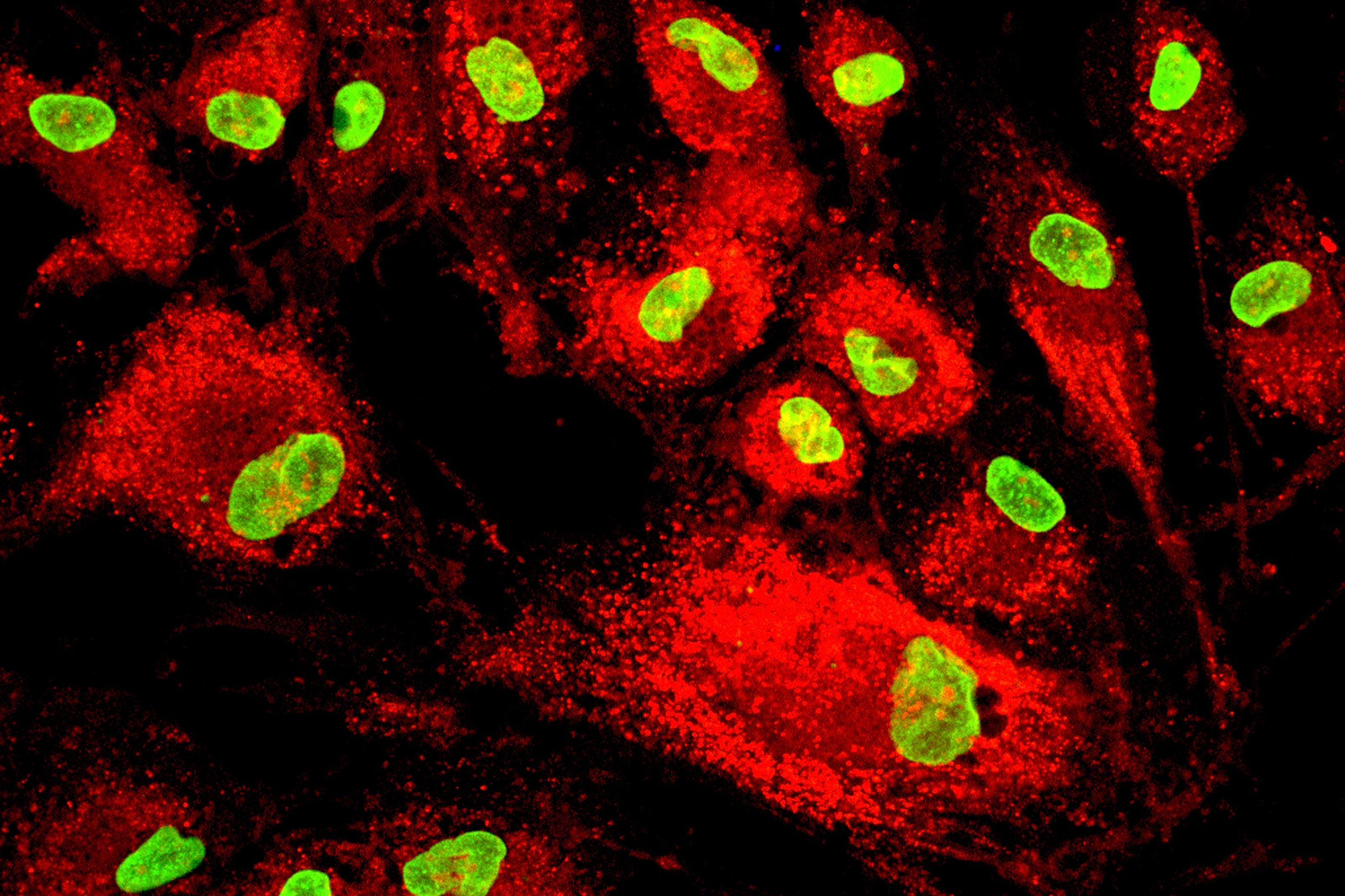
We obtain the stem cells from the patient’s own fat tissue. Stem cells from fat tissue are called adipose-derived stem cells (ADSCs or adMSCs). Fat tissue contains MSC in relatively high numbers. In the laboratory the non-cellular parts of the tissue are removed enzymatically or mechanically. The cell mixture that remains is called the stromal vascular fraction (SVF). Only about 1 to 10 % of its cells are MSC and progenitor cells.
Which Stem Cells Do We Culture?
We culture mesenchymal stem cells (MSC) from the SVF of the patient’s own fat tissue. In our clean-room laboratory this small number of cells is grown and multiplied. The result is a pure culture of 90 to 98 % MSC. This gives far more stem cells than BMC, which is not multiplied. During culture the cells release substances that research describes as possibly regenerative and anti-inflammatory. We collect and concentrate these substances for treatment. ANOVA multiplies the cells only within the careful limits recognised in research. In contrast to other clinics and manufacturers our cell numbers are lower but the cells are “less expanded” increasing the potential for unchanged secretomes.
How MSEC Is Produced
The production of MSEC takes three steps, followed by the application. The treatment can last from a few weeks to two years.
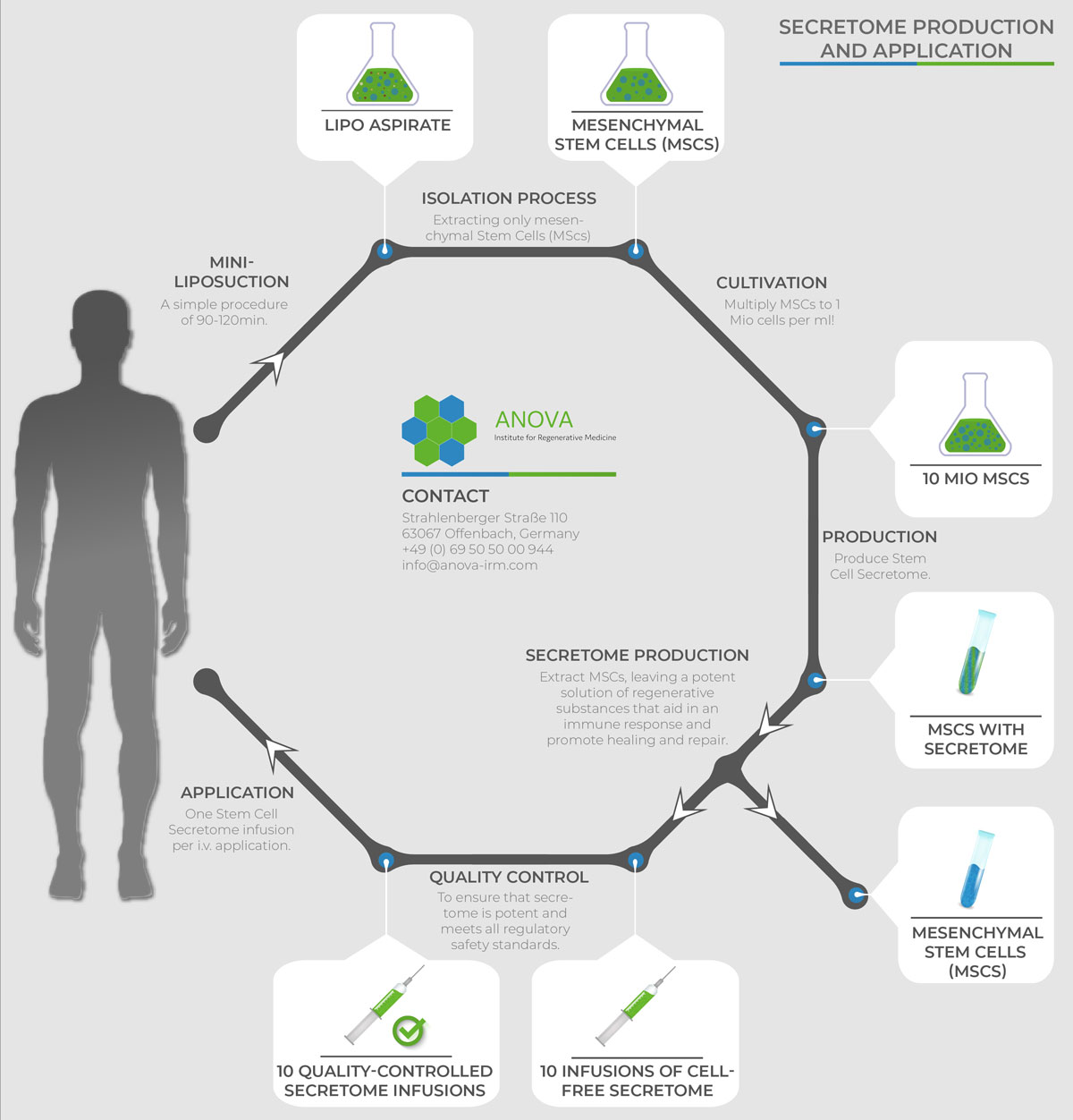
Step 1 — Collecting the fat tissue
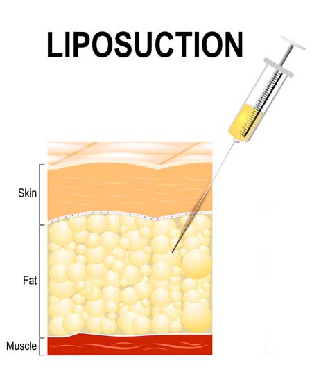
We collect a small amount of fat tissue from under the skin in a mini-liposuction. ANOVA uses the so-called nutational infrasonic liposuction with the Euromi Lipomatic® system. This technique is designed to be gentle on the tissue and to keep the cells in it viable. The tissue is processed immediately in our clean room under defined quality controls.
Step 2 — Culturing the cells and producing the secretome
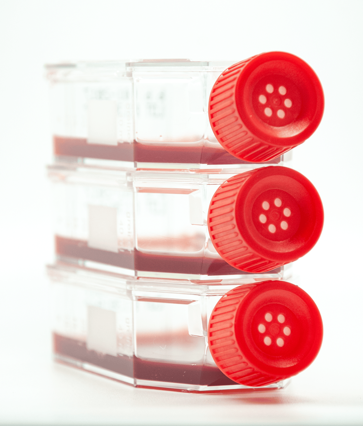
In the clean-room laboratory the SVF is isolated from the tissue by enzymatic digestion. SVF is a cell mixture and contains low numbers of stem cells. The MSC it contains are therefore multiplied until the required number is reached, and their purity rises to 90 to 98 % (see Which stem cells do we culture?). ANOVA multiplies the cells only within the careful limits recognised in research, because their potential effect declines the more they are multiplied [44]. As a last step the cell-free secretome is produced and frozen.
Step 3 — Quality control, release and storage
Every batch is then fully tested before release (see Quality control). The released secretome is stored at −80 °C and keeps for up to two years. One collection gives up to ten doses.
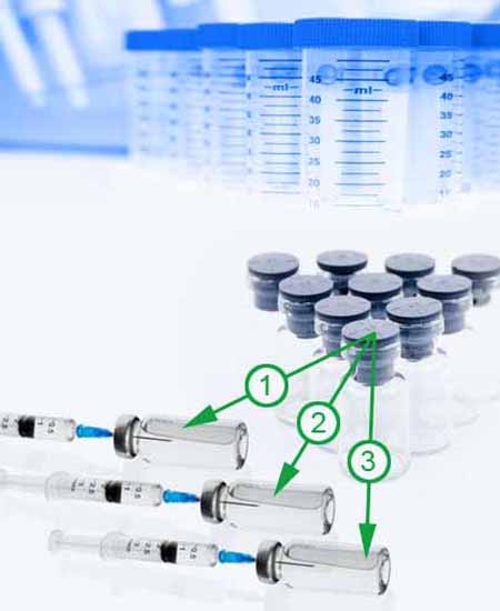
Step 4 — Application
The first dose can be given four to six weeks after the liposuction. During this time our laboratory produces and tests the product. The doses are given into affected sites or intravenously where appropriate (see Routes of administration).
Ten doses allow a treatment plan with several applications over up to two years for chronic diseases or shorter time intervals for higher dosage per time.
Quality Control and Release of MSEC
Every batch of MSC secretome is tested before it is released for use. Officially, MSEC is regulated under GFP. Even so, ANOVA applies the higher GMP standards to all testing. All tests are approved and inspected by the Paul-Ehrlich-Institut (PEI) and the Hessian State Office for Health and Care (HLfGP). All methods are validated and, where applicable, follow the European Pharmacopoeia (Ph. Eur.). Every release is documented. All release tests are completed before the first dose is given.
| Release test | Every batch | Before release / before application | Standard |
|---|---|---|---|
| Viable cell count per dose | Yes | Yes | EMEA/CHMP/410869/2006; methods according to Ph. Eur. 2.7.29 |
| Viability | Yes | Yes | EMEA/CHMP/410869/2006; methods according to Ph. Eur. 2.7.29 |
| MSC identity | Yes | Yes | internal SOP |
| Microbiological control (sterility) | Yes | Yes | Ph. Eur. 2.6.27 |
| Endotoxin content | Yes | Yes | Ph. Eur. 2.6.14 |
| Mycoplasma | Yes | Yes | Ph. Eur. 2.6.7 |
| Particle content (approximate EV content) | Yes | Yes | internal SOP |
| Total population doublings | Yes | Yes | internal SOP |
| Total culture time | Yes | Yes | internal SOP |
| Final product volume | Yes | Yes | internal SOP |
| Number of final product vials | Yes | Yes | internal SOP |
Some tests are not needed for every batch. The differentiation potential of the MSC and the EV content and identity were tested during product characterisation. The production process ensures that the final product is cell-free. No karyotyping is required for MSEC, because MSEC is cell-free and therefore cannot contain cancer cells. At no point are cells frozen and taken back into culture.
If a batch does not comply with the set product specifications, it is not released.
Routes of Administration
MSC secretome is not given the same way for every condition. The route is chosen so that the preparation reaches the affected area. It is decided with the treating physician as part of the individual benefit-risk assessment. Injection is done under local anaesthesia where necessary or under slight sedation by our anaesthesiologist when joints are the target.
Intrathecal — neurodegenerative disease and spinal cord injury
For neurodegenerative diseases and spinal cord injury, MSEC is given intrathecally. This means a lumbar puncture into the cerebrospinal fluid. The reason is anatomical. The cerebrospinal fluid is a small closed space in direct contact with the spinal cord and the brain. The preparation therefore reaches the affected tissue directly instead of spreading through the whole body. Whether the secretome can reach the brain from the blood is discussed on the page does the secretome pass the blood-brain barrier?. If the anatomy makes a normal puncture difficult, the injection can be guided by CT (see diagnostics at ANOVA IRM). A lumbar puncture is more invasive than an infusion and has its own risks. These are explained in the benefit-risk assessment of intrathecal administration.
Intracavernosal — erectile dysfunction
For erectile dysfunction, MSEC is injected directly into the erectile tissue of the penis (corpora cavernosa). The preparation is placed in the affected tissue rather than in the bloodstream.
Systemic administration
For systemic indications such as anti-ageing, it is given as an infusion. The route for each condition is described on the relevant disease page.
Individual Treatment Plans and Cost
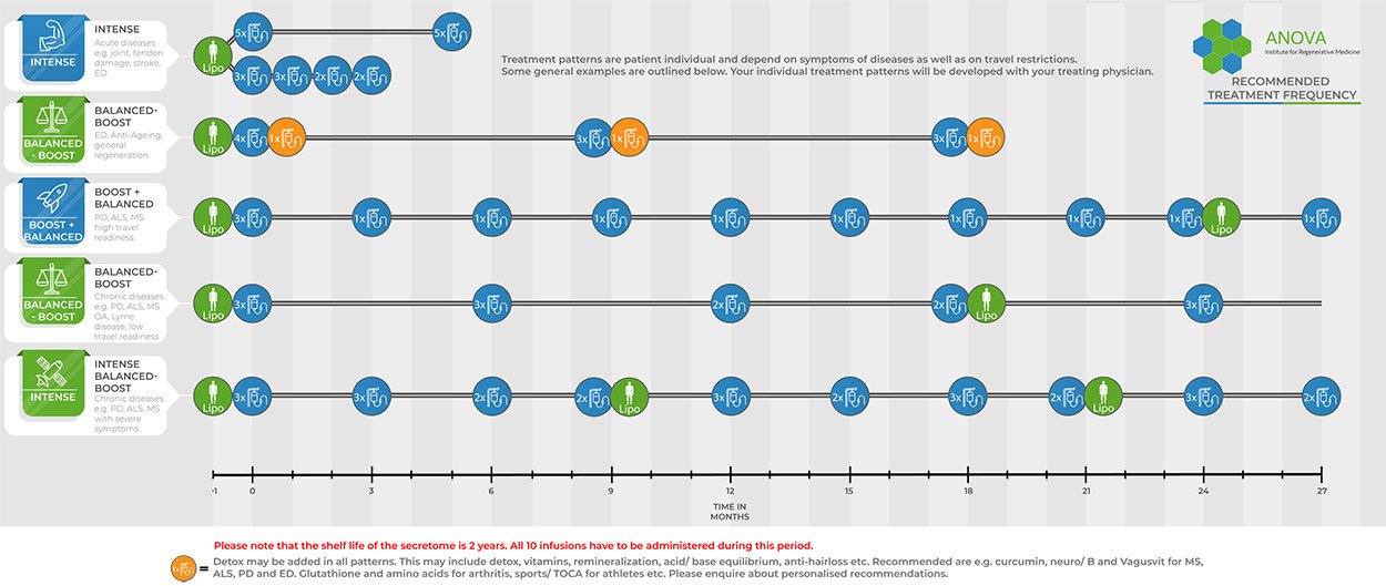
Each treatment plan is drawn up individually. It takes into account the disease, its stage and your personal situation. One standard mini-liposuction gives 10 doses. The secretome keeps for up to two years. This allows a planned course of treatment instead of a single treatment at one point in time.
The schedule is set individually for each patient, for example:
- Intense: In acute diseases the applications usually follow each other quickly.
- Boost and balanced: For patients from Germany and nearby countries such as Austria, Switzerland, Italy, the Netherlands, the UK, France or Spain, several trips are usually easy and inexpensive. Their plan usually starts with a series of doses (boost), followed by a single dose every 4 to 6 weeks (balanced).
- Fewer, longer visits: Patients from further away, for example from the USA, Canada, South America, Brazil, the Middle East, Saudi Arabia, Oman or Russia, usually receive a double dose every 3 months. This reduces the number of trips.
Cost
MSEC treatment with a single liposuction and 10 doses costs approximately €20,000 to €36,000 with intravenous application. It is billed according to the German Medical Fee Schedule (GOÄ). The base package costs approximately €20,000 and includes the mini-liposuction and the first three MSEC doses. Each further dose costs approximately €2,250 to €2,350, depending on the application route. Intrathecal application costs €100 more per dose than intravenous application. The total depends on how many of the 10 doses are used, on additional examinations and on your sedation or anaesthesia preferences. You receive an individual written cost estimate before you decide on treatment. Travel, accommodation and other personal expenses are not included.
For chronic diseases a double liposuction can be recommended. It gives 20 doses, which can be used over the current shelf life of two years from a single collection. The cost is correspondingly higher and is shown in your individual estimate.
Experimental treatment is generally self-funded. Statutory and private health insurers do not cover these costs. You can check possible reimbursement with your insurer, but you should expect to pay the costs yourself. All prices are subject to change.
Who Qualifies for MSEC Treatment, and What Are the Requirements?
We treat patients whose condition is one for which the literature, research and study data suggest that a clear benefit may be possible. You must be medically fit for the brief sedation required to collect fat tissue. Every treatment recommendation follows a full clinical evaluation of your medical history and current findings.
What Are the Contraindications for MSEC Treatment?
Our treatments are experimental. We treat only people for whom, after medical assessment, we consider the benefit-risk balance to support treatment.
Applies to all ANOVA treatments:
- Active cancer within the last two years
- Under the age of legal majority
- Pregnancy or breastfeeding
- Active infectious disease (hepatitis A, B, C, HIV, syphilis or other)
Further condition-specific exclusion criteria are set out on the relevant disease page.
What Does MSEC Therapy at ANOVA IRM Involve, Step by Step?
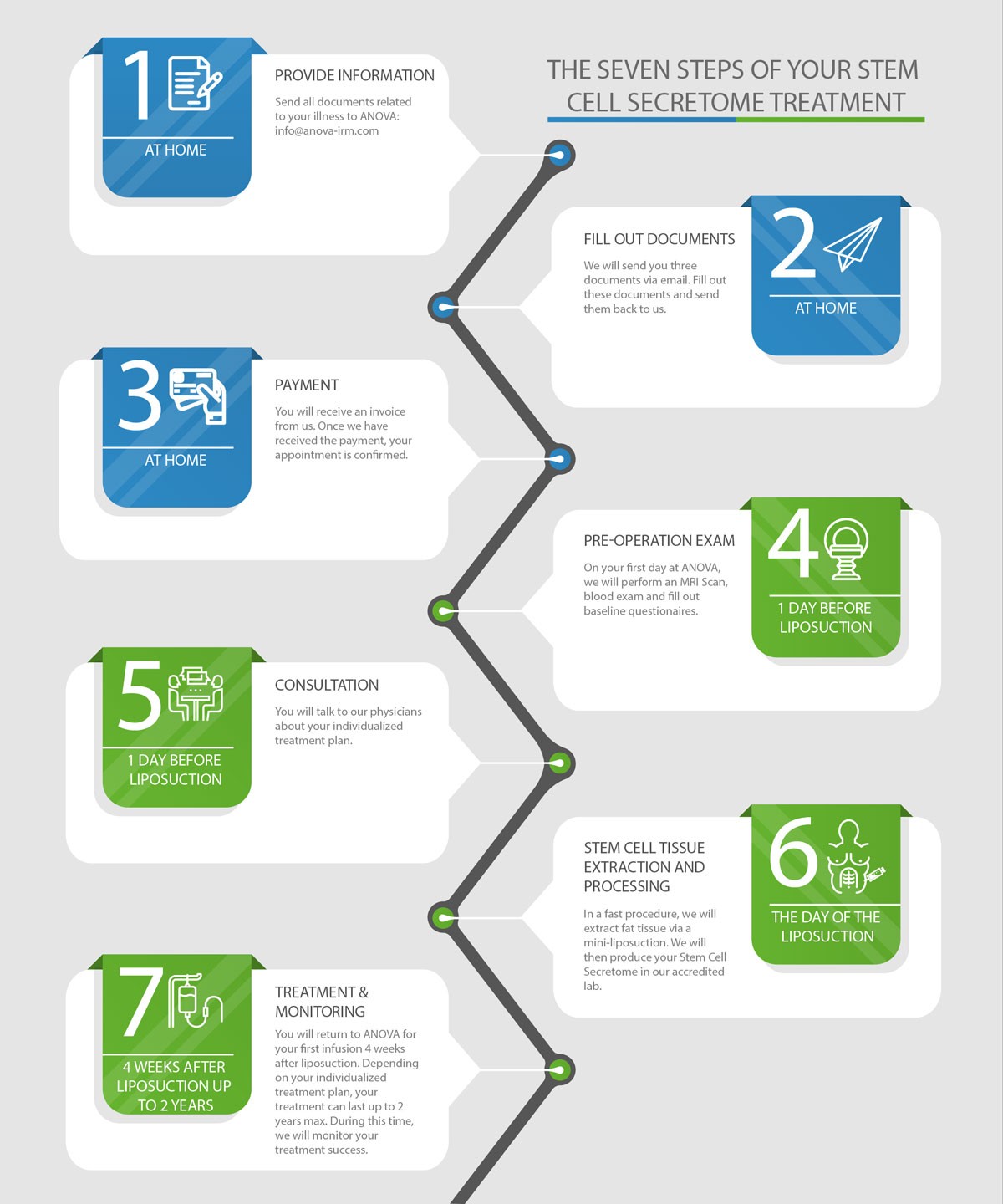
Your therapy at ANOVA IRM starts with your first free contact and enquiry. We check whether our therapies are suitable for your condition and whether you are eligible as a donor. You then receive a cost estimate and a treatment contract. Appointments with our physicians can take place virtually or in person.
- 01
Initial evaluation
Remote, by phone or video — typically 2 weeks to a few months
- 02
Preliminary screening
In your home country, to avoid an unnecessary journey
- 03
First visit — bloodwork and tissue collection
Two-day outpatient visit to Offenbach: bloodwork first, then — if the results are in order — mini-liposuction under brief sedation
- 04
Production and quality control
4 to 6 weeks; yields 10 doses per production cycle
- 05
Storage
Up to 2 years, allowing an extended personalised schedule
- 06
Application schedule
Set individually, for example two doses at the start, then one dose every 4–6 weeks, or two doses every 3 months
What Screening Is Needed Before You Travel?
Before you travel, a preliminary screening in your home country helps you avoid an unnecessary journey. It is usually a blood test for medical or infectious-disease factors that would prevent treatment.
What Happens at Your First Visit to Offenbach, Germany?
The first treatment visit takes about two days in Offenbach, on an outpatient basis. It begins with bloodwork at ANOVA IRM. If the results are in order, a small amount of abdominal fat tissue is collected by mini-liposuction under brief sedation. From this point to the first secretome application usually takes four to six weeks.
ANOVA IRM carries out the full donor screening required under EU GMP and the German AMWHV (Arzneimittel- und Wirkstoffherstellungsverordnung). Your blood is analysed in a contract laboratory that holds the corresponding GMP/GFP licences. This is why you arrive one day before the treatment. ANOVA IRM treats only patients without an infectious disease.
How Is Your MSEC Produced and Quality-Controlled?
The production and testing of your MSEC are described above in How MSEC is produced and Quality control.
How Often Is MSEC Applied?
The schedule is set individually for each patient. Examples are given above in Individual treatment plans.
Frequently Asked Questions — Production and Treatment
How is secretome produced? What tissue is used for secretome production?
The secretome is produced from your own mesenchymal stem cells (MSC). They come from a small amount of abdominal fat tissue collected by mini-liposuction. In our clean-room laboratory the MSC are isolated, multiplied to a purity of 90 to 98 % and stimulated to release the secretome. Each batch is tested before release. Details are given in MSC isolated from fat tissue, How MSEC is produced and Quality control.
Does ANOVA use donor cells?
No. The ANOVA secretome is autologous, which means it is made from your own cells. Products from unrelated donors carry immunological and infection risks that do not arise with an autologous preparation.
What are the side effects of secretome/exosome treatment?
The side effects are usually minimal. They come mainly from the mini-liposuction and the application: soreness, bruising or swelling where the fat tissue was collected, and pain or, rarely, bleeding or infection at the injection site. After a lumbar puncture for intrathecal application, a temporary headache can occur. To date, no serious adverse event has been caused by the product. Your doctor explains these risks to you before treatment.
Can secretome/exosomes cause cancer?
No cultured cells are transferred, so the risk that is specific to cell products does not apply. No karyotyping is required for the ANOVA secretome, because it is cell-free and therefore cannot contain cancer cells. For MSC, the literature reports genetic changes in long-term cultures but no confirmed change into cancer cells [42],[43]. Patients with active cancer within the last two years are not treated. More on tumour risk in cell products and on how safe ANOVA MSC secretome is. ANOVA IRM uses the whole secretome, not only exosomes.
How often do I need to travel to Germany for secretome/exosome treatment?
Usually several times, depending on your treatment plan. We use secretome for chronic diseases, and no stem cell treatment offers a cure. Treatment is therefore planned as a longer course with several applications. The first visit takes about two days in Offenbach, less than 20 minutes from Frankfurt Airport, and includes the collection of fat tissue. The first application is possible four to six weeks later. If you live close by, you usually come for a single dose every 4 to 6 weeks. If you live further away, you usually come for a double dose every 3 months, which means about five visits over 15 months for 10 doses. The initial evaluation can take place remotely by phone or video. ANOVA IRM uses the whole secretome, not only exosomes.
What research says about MSC and their secretome, how MSEC may work in each condition, and all references.
You are hereThe Science of Secretome and Exosomes
What Is Research Investigating About the Potential of Mesenchymal Stem Cells?
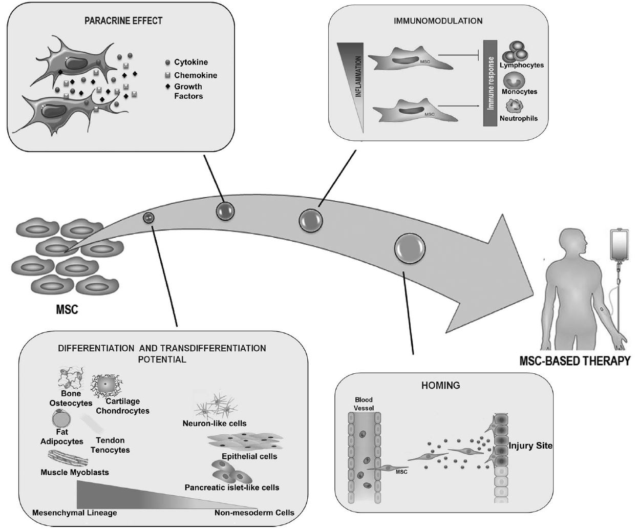
Research is investigating four properties of MSC:
- They can develop into various cell types in the culture dish. This is not thought to be how they work as a treatment, because in the body stem cells do not differentiate into tissue cells.
- They release paracrine factors that are being studied for a possible role in healing and regeneration of surrounding cells.
- Laboratory studies have observed possible effects on inflammation and immune regulation.
- Preclinical models describe how MSC migrate towards sites of injury and influence the cells there.
SVF or Cultured MSC?
SVF itself has been examined in early clinical research for some conditions. These findings are still preliminary, and the number of MSC in SVF is limited. We reject SVF as a product, because it contains only few stem cells compared with cultured cells: 1 to 10 % MSC in SVF against 90 to 98 % in an expanded culture. MSC can be multiplied in the laboratory relatively easily. However, this changes the risk profile and makes clinical use much more complex. For this reason the multiplication of cells is highly restricted in all developed countries. External laboratory studies suggest that a high number of cells may not be the decisive factor. What may matter more is how vital the cells are and which substances they release. This knowledge led the scientists at ANOVA IRM to a different approach: the stem cell secretome. It contains active components such as growth factors and cytokines. Laboratory research is studying them as possible mediators of the regenerative effects seen in preclinical models.
Are MSC Studies Evidence for MSEC? From Cells to Cell-Free
A frequent objection is that studies with mesenchymal stem cells do not prove that MSEC does the same. That is correct. MSC studies do not prove that MSEC is effective. They are still relevant, and the reason lies in how MSC work. The field itself has taken this path, from cells towards cell-free preparations.
The mesenchymal stem cell secretome, or MSC secretome, is also called the mesenchymal stromal cell secretome. Mesenchymal stem cells and mesenchymal stromal cells are two names for the same cells (MSC); the International Society for Cell & Gene Therapy recommends “stromal” for cells grown in the laboratory [29],[30]. MSEC is therefore a mesenchymal stromal cell secretome made from your own cells.
MSC do not stay in the body for long. After an intravenous infusion most of them are trapped in the lungs and disappear within days. Long-term engraftment in humans is minimal [1],[5],[6].
Their effects persist through released factors. MSC act mainly through substances they release (paracrine action; [31]). When a single released anti-inflammatory protein (TSG-6) was switched off, the benefit of MSC after a heart attack disappeared. The protein alone reduced inflammation and the size of the infarct [5].
Cell-free products can reproduce MSC effects. In a direct comparison after stroke, extracellular vesicles from MSC were not inferior to the cells themselves [80]. MSC-conditioned medium reduced infarct size in a large-animal model [139]. In a metabolic osteoarthritis model the vesicles performed better than the cells [112].
Hence the move to cell-free. Reviews now describe the secretome as a cell-free approach to regeneration [14],[15],[16]. If MSC act largely through their secretome, a secretome from the same cell type delivers these mechanisms without transferring cultured cells.
Where the limits are. The secretome is not identical to living MSC. Some effects may depend on living or dying cells interacting with the recipient’s immune system [7]. The composition also depends on the manufacturing process. To our knowledge no randomised clinical trial has directly compared MSC with their secretome in patients. MSC studies are therefore a scientific rationale and indirect evidence, not proof that MSEC works. This is why MSEC remains experimental.
The Science of Secretome
Laboratory, animal and early clinical studies have examined how MSC and their secretome act. They point to several mechanisms. Most of the data come from animal models. Human studies with cell-free MSC products are still few and small. We offer MSEC only where these data suggest that a clear benefit may be possible.
Anti-inflammatory action and immune balance
- In mice after a heart attack, MSC released the anti-inflammatory protein TSG-6. When TSG-6 was switched off, the benefit disappeared. The protein alone reduced inflammation and infarct size [5].
- Exosomes from MSC made immune cells release anti-inflammatory signals and increased regulatory T cells, which calm the immune response [3].
- Exosomes from MSC were more immunosuppressive than larger microparticles in a mouse model of inflammatory arthritis [104].
- In a single case report, a patient with therapy-resistant graft-versus-host disease was treated with MSC-derived vesicles as an individual treatment attempt; the authors report clinical improvement [137].
Neuroprotection and neuronal survival
- In rats with Parkinson's disease, the secretome of human MSC increased the number of dopamine-producing neurons and partly restored motor performance [66].
- In the same model, the secretome of bone marrow MSC protected dopamine-producing neurons better than transplanting the cells themselves [67].
- After stroke in mice, vesicles from MSC were not inferior to the cells. They protected brain tissue in the long term and improved motor coordination [80].
- In ALS mice (SOD1-G93A), repeated doses of exosomes from adipose-derived stromal cells improved motor performance and protected motor neurons, neuromuscular junctions and muscle [60].
Less glial scarring
- After spinal cord injury in rats, exosomes from bone marrow MSC reduced glial scar formation, lesion size and nerve cell death. They suppressed harmful (A1) astrocytes and improved movement [91].
- In a second rat study, MSC and their exosomes reduced A1 astrocytes and inflammatory signals. Given into a vein, the exosomes worked about as well as the cells [92].
- In a model of progressive MS, vesicles from human MSC reduced the activation of astrocytes and microglia in the brain [76].
Myelin repair
- In MS models, the secretome of human MSC reduced deficits and promoted myelin-forming cells. This depended on hepatocyte growth factor (HGF), which alone sped up remyelination [77].
- Vesicles from human adipose MSC improved motor deficits, reduced brain atrophy and increased myelin proteins in a model of progressive MS [76].
- Vesicles from placental MSC increased myelination of the spinal cord in mice with an MS-like disease [78].
Inflammation causes pain — pain reduction
- In rats with knee osteoarthritis, exosomes from bone marrow MSC reduced pain sensitivity and inflammatory pain markers and preserved cartilage [116].
- In mice with osteoarthritis, the secretome of MSC reduced pain and prevented cartilage damage [120].
- In rats with nerve-injury pain, exosomes from umbilical cord MSC given intrathecally prevented and reversed pain hypersensitivity. They lowered inflammatory signals and raised the nerve growth factors BDNF and GDNF [4].
Less fibrosis in the erectile tissue
- In rats with cavernous nerve injury, exosomes from MSC improved erectile function, improved the ratio of smooth muscle to collagen and reduced cell death, with an effect similar to the cells [96].
- In diabetic rats, exosomes from adipose-derived stem cells improved erectile function, increased smooth muscle relative to collagen and reduced cell death in the erectile tissue [97].
- In a second study in diabetic rats, these exosomes restored erectile function and reduced fibrosis of the erectile tissue [98].
- A cell-free lysate of adipose-derived stem cells improved erectile function in rats to a similar extent as the cells [100].
New blood vessels
- The conditioned medium of MSC alone increased the growth of blood vessel cells and improved blood flow in mice with limb ischaemia [145].
- Vesicles from bone marrow MSC increased new blood vessels and blood flow in rats after a heart attack and reduced infarct size [146].
Cartilage protection
- Exosomes and microparticles from MSC protected cartilage and bone in mice with osteoarthritis as well as the cells did [117].
- In rats, exosomes from MSC restored cartilage and the underlying bone in joint defects, while untreated defects formed only fibrous tissue [118],[119].
Senescence and ageing
- Vesicles from young MSC reduced markers of cell ageing and extended the period of good health in two mouse models of ageing [132].
- Vesicles from umbilical cord MSC rejuvenated aged stem cells and slowed age-related bone and kidney degeneration in old mice [131].
First clinical studies with cell-free MSC products
- Chronic kidney disease: In a small randomised pilot study with 40 patients, vesicles from umbilical cord MSC caused no significant side effects over one year, and kidney function improved in the treated group [134]. The study was small and single-centre.
- Alzheimer's disease: In a phase 1/2 study with 9 patients, exosomes from adipose MSC given through the nose caused no adverse events; memory scores improved in the medium-dose group. There was no control group [83].
- COVID-19 respiratory failure: In a small external randomised, placebo-controlled phase 2 trial with 102 patients, vesicles from bone marrow MSC caused no treatment-related adverse events. The main goal, lower 60-day mortality, was not reached [133]. Earlier uncontrolled studies had reported good tolerability [135],[138].
- Stroke: In a pilot study with 5 patients, exosomes from placental MSC injected into brain tissue caused no serious adverse events. The study examined safety only [79].
- Hair loss: In a retrospective case series with 27 women, the conditioned medium of adipose stem cells increased hair density and thickness without severe side effects [128].
These studies used products from different sources and manufacturing processes, often from donor cells. Their results cannot be transferred directly to MSEC. They show which mechanisms research is investigating and that cell-free MSC products have so far been well tolerated in small clinical studies.
How MSEC May Work in Each Condition
The assumed mechanism of MSEC is similar in all conditions. The released factors are thought to influence the environment of damaged or inflamed cells. Which processes are targeted differs from disease to disease. For each condition we summarise the hypothesis, the laboratory and animal data and the clinical data. None of these mechanisms has been proven in patients for MSEC. ANOVA IRM has not yet tested MSEC in a clinical study.
ALS
Hypothesis. ALS is thought to start in the motor neurons, while supporting cells such as microglia and astrocytes drive the progression through inflammation. The soluble factors of MSEC are thought to influence this environment rather than the motor neuron itself.
Laboratory and animal data. In ALS mice, exosomes from adipose-derived stromal cells improved motor performance and protected motor neurons, neuromuscular junctions and muscle [60]. In the same mouse model, blocking three inflammatory factors released by microglia markedly extended survival [61].
Clinical data. Small phase 1 and phase 1/2 studies with MSC products given intrathecally mainly examined safety and reported acceptable tolerability [57],[58]. In two phase 3 trials with MSC products (NurOwn and NeuroNata-R), the main goal was not reached in the full study population [55],[56]. For NeuroNata-R this result has so far only been reported by the manufacturer. NeuroNata-R has a conditional approval in South Korea [58],[59].
Limitation. Anti-inflammatory approaches have so far had only modest effects in ALS. The aim is to slow progression, not to cure. More in the scientific rationale for ALS.
Parkinson's disease
Hypothesis. The dopamine-producing neurons die gradually, surrounded by an inflammatory environment. Neurotrophic factors such as BDNF and GDNF and other factors of the secretome are thought to dampen inflammation and support the remaining neurons.
Laboratory and animal data. A systematic review of 13 animal studies found that vesicles from MSC improved motor tests, lowered inflammatory markers and preserved dopamine-producing neurons [68]. In rats, the secretome of human MSC increased the number of these neurons and improved motor performance [66]. It protected them better than the cells themselves [67].
Clinical data. Small clinical studies tested MSC cells, not secretome. In an open study with seven patients, three improved steadily; there was no control group [65]. A small external phase 1 trial with 20 patients reported no serious reactions [64]. A small external randomised phase 2 trial with 45 patients gave mixed results [62]. In a second small randomised phase 2 trial with 24 patients, a clear effect could not yet be demonstrated [63].
Limitation. Neurons already lost are not expected to be replaced. More in the scientific rationale for Parkinson's disease.
Multiple sclerosis
Hypothesis. An overactive immune system attacks the myelin sheath. The secretome is thought to influence immune activity, inflammatory signalling and the survival of the cells involved.
Laboratory and animal data. In MS models, the secretome of MSC reduced deficits and promoted myelin repair through hepatocyte growth factor [77]. Vesicles from MSC improved motor function, reduced brain atrophy and increased myelin proteins [76],[78].
Clinical data. Small phase 1 and phase 2 studies with MSC cells mainly examined safety and reported acceptable tolerability [71],[74]. In small external randomised placebo-controlled phase 2 trials, a clear clinical benefit could not yet be demonstrated [69],[70].
Limitation. The cause of MS remains unknown. More in the scientific rationale for MS.
Spinal cord injury
Hypothesis. Inflammation, glial scarring and poor blood supply limit the surviving nerve fibres. The secretome, given into the cerebrospinal fluid, is thought to make this environment more permissive for the remaining pathways.
Laboratory and animal data. In rats, exosomes from MSC reduced glial scarring, lesion size and nerve cell death and improved movement [91],[92]. Grafted MSC improved recovery through growth factors and new blood vessels [94].
Clinical data. Reviews of mostly small and uncontrolled MSC studies describe acceptable short-term tolerability and improvements in some patients [89],[90]. Meta-analyses of the controlled studies could not yet show a clear benefit [87],[88]. In a randomised placebo-controlled phase 1/2 trial with intrathecal MSC, a significant difference from placebo could not yet be demonstrated [86].
Limitation. The combination with HAL training has not yet been compared with HAL alone. More in the scientific rationale for SCI.
Erectile dysfunction
Hypothesis. The released factors are studied for possible effects on the blood vessel lining, the smooth muscle cells and the nerves of the erectile tissue. Possible goals are slower fibrosis of the erectile tissue and less chronic inflammation.
Laboratory and animal data. In rats with nerve or artery injury and in diabetic rats, exosomes from MSC or adipose-derived stem cells improved erectile function. They also increased smooth muscle relative to collagen and reduced cell death and fibrosis [96],[97],[98],[99]. A cell-free lysate worked about as well as the cells [100].
Clinical data. There is no clinical study with secretome in ED. In 17 men after prostate removal, a single injection of fresh fat-derived regenerative cells was well tolerated, and 8 were able to have intercourse again; there was no control group [95].
Limitation. The human data come from living cells, not from a secretome. More in the scientific rationale for ED.
Rheumatoid arthritis
Hypothesis. An immune over-reaction keeps the joint lining inflamed. The secretome is thought to balance immune responses, dampen acute inflammation and support repair of joint tissue.
Laboratory and animal data. Exosomes from MSC inhibited T cells and increased regulatory T cells. In mice with arthritis they reduced the signs of inflammation more than larger microparticles [104].
Clinical data. All clinical studies used MSC cells. In a phase 1/2 study, 15 patients with RA of the knee received cells into the joint and 15 received saline. Pain and function improved more with the cells, but not beyond 12 months [102]. In a controlled study with 172 patients, umbilical cord MSC in a vein lowered disease activity for three to six months [103]. In a placebo-controlled phase 1b/2a study with 53 patients, a clear benefit could not yet be demonstrated [101].
Limitation. The clinical data come from living cells. More in the scientific rationale for RA.
Joint and back pain
Hypothesis. For osteoarthritis, joint injuries and back pain, BMC is the standard product at ANOVA IRM. External laboratory, animal and small clinical studies with cultured MSC or their secretome point in the same direction: less inflammation, less pain and protection of cartilage and disc tissue.
Laboratory and animal data. In animals with osteoarthritis, the secretome or exosomes of MSC reduced pain and protected cartilage [116],[117],[120]. In laboratory studies, stem cells exchanged vesicles with disc cells [124].
Clinical data. In a small phase 1/2 study, MSC injected into knees with osteoarthritis improved function and reduced cartilage defects [107]. After partial meniscus removal, donor MSC were followed by meniscus regrowth in some patients [108]. In the disc, small studies reported less pain and disability [110],[111]. In a phase 3 trial with 404 patients and donor precursor cells, the combined main goal could not yet be demonstrated [106].
Limitation. These products differ from both BMC and MSEC. More on the pages for osteoarthritis and back pain.
Anti-ageing
Hypothesis. With age, senescent cells and chronic low-grade inflammation increase, while repair signals decline. The secretome is thought to reduce inflammatory signalling, alone or after the removal of senescent cells.
Laboratory and animal data. Vesicles from young MSC reduced markers of cell ageing and extended the period of good health in two mouse models of ageing [132]. Vesicles from umbilical cord MSC slowed age-related bone and kidney degeneration in old mice [131].
Clinical data. There is no clinical study of MSEC or other MSC secretomes on ageing. A change in blood markers alone would not prove a longer healthy life.
Limitation. All data come from animals. More in the scientific rationale for anti-ageing.
Frequently Asked Questions — Evidence
Can MSC secretome cure diseases?
No. No stem cell treatment and no treatment derived from stem cells offers a cure anywhere in the world. The only exception is gene therapy for a small number of specific genetic disorders. Secretome treatment is investigated for its potential to slow progression and support remaining tissue, not to cure. Anyone who offers you a cure is not describing the state of the evidence.
Is secretome less effective than MSC therapy?
In several animal studies it was not. When cell-free MSC products were compared directly with the cells, they worked about as well after stroke [80] after spinal cord injury [92] in erectile dysfunction [96],[100] and in osteoarthritis [117]. In a mouse model of ageing, vesicles from young MSC extended lifespan in a similar way to the young cells themselves [132]. In Parkinson’s disease [67] and in a metabolic osteoarthritis model [112] the cell-free product was even more effective. These results support the idea that MSC act largely through their secretome. In patients, however, no randomised trial has yet compared the two. More in The science of secretome.
Is MSC literature proof that secretome/exosomes are effective?
No. MSC studies provide a scientific rationale and indirect evidence, not proof. Proof of efficacy for a specific product in a specific condition requires a randomised controlled phase 3 trial with that product. No such trial exists for the ANOVA secretome. Even for living MSC, randomised trials show mixed results. In multiple sclerosis, for example, a randomised trial of intravenous MSC showed no benefit over placebo [69]. More in Are MSC studies evidence for MSEC? and on the page unproven therapies.
Important Pages
Questions people usually ask next. Each is answered in full on its own page.
- Who are we?
- Is ANOVA legal?
- Is stem cell therapy safe?
- What is BMC (Bone Marrow Concentrate)?
- What are stem cells?
- What should I ask when looking for a stem cell treatment?
- Which conditions do we treat?
- Benefit-risk assessment of intrathecal administration
- Does the secretome pass the blood–brain barrier?
Affiliations of ANOVA
ANOVA IRM shares its premises in Offenbach with two further institutions owned by Dr. Stehling: the Institut für Bildgebende Diagnostik (IBDO), providing MRI and CT imaging, and the VITUS PRIVATKLINIK Prof. Dr. Stehling GmbH. Because imaging is carried out in the same building, diagnostics — including CT-guided procedures such as intrathecal applications — can be performed in-house rather than referred elsewhere. See diagnostics at ANOVA IRM.
References and Literature — MSC Secretome
This page is the reference home for secretome, exosome and extracellular vesicle literature. Other pages link here rather than repeating the list.
General — secretome, extracellular vesicles and MSC
Other clinical studies and reviews of clinical studies
- [1] von Bahr L, Batsis I, Moll G, et al. Analysis of tissues following mesenchymal stromal cell therapy in humans indicates limited long-term engraftment and no ectopic tissue formation. Stem Cells. 2012;30(7):1575-8. doi:10.1002/stem.1118. PMID: 22553154.
Animal studies
- [2] Mitchell R, Mellows B, Sheard J, et al. Secretome of adipose-derived mesenchymal stem cells promotes skeletal muscle regeneration through synergistic action of extracellular vesicle cargo and soluble proteins. Stem Cell Res Ther. 2019;10(1):116. doi:10.1186/s13287-019-1213-1. PMID: 30953537.
- [3] Zhang B, Yin Y, Lai RC, et al. Mesenchymal stem cells secrete immunologically active exosomes. Stem Cells Dev. 2014;23(11):1233–1244. doi:10.1089/scd.2013.0479. PMID: 24367916.
- [4] Shiue SJ, Rau RH, Shiue HS, et al. Mesenchymal stem cell exosomes as a cell-free therapy for nerve injury-induced pain in rats. Pain. 2019;160(1):210–223. doi:10.1097/j.pain.0000000000001395. PMID: 30188455.
- [5] Lee RH, Pulin AA, Seo MJ, et al. Intravenous hMSCs improve myocardial infarction in mice because cells embolized in lung are activated to secrete the anti-inflammatory protein TSG-6. Cell Stem Cell. 2009;5(1):54-63. doi:10.1016/j.stem.2009.05.003. PMID: 19570514.
- [6] Eggenhofer E, Benseler V, Kroemer A, et al. Mesenchymal stem cells are short-lived and do not migrate beyond the lungs after intravenous infusion. Front Immunol. 2012;3:297. doi:10.3389/fimmu.2012.00297. PMID: 23056000.
- [7] Galleu A, Riffo-Vasquez Y, Trento C, et al. Apoptosis in mesenchymal stromal cells induces in vivo recipient-mediated immunomodulation. Sci Transl Med. 2017;9(416):eaam7828. doi:10.1126/scitranslmed.aam7828. PMID: 29141887.
Laboratory studies
- [8] Sze SK, de Kleijn DP, Lai RC, et al. Elucidating the secretion proteome of human embryonic stem cell-derived mesenchymal stem cells. Mol Cell Proteomics. 2007;6(10):1680-9. doi:10.1074/mcp.M600393-MCP200. PMID: 17565974.
- [9] Lee MJ, Kim J, Kim MY, et al. Proteomic analysis of tumor necrosis factor-alpha-induced secretome of human adipose tissue-derived mesenchymal stem cells. J Proteome Res. 2010;9(4):1754-62. doi:10.1021/pr900898n. PMID: 20184379.
- [10] Chen TS, Lai RC, Lee MM, et al. Mesenchymal stem cell secretes microparticles enriched in pre-microRNAs. Nucleic Acids Res. 2010;38(1):215-24. doi:10.1093/nar/gkp857. PMID: 19850715.
- [11] Lopez-Verrilli MA, Caviedes A, Cabrera A, et al. Mesenchymal stem cell-derived exosomes from different sources selectively promote neuritic outgrowth. Neuroscience. 2016;320:129-39. doi:10.1016/j.neuroscience.2016.01.061. PMID: 26851773.
- [12] Wilkins A, Kemp K, Ginty M, et al. Human bone marrow-derived mesenchymal stem cells secrete brain-derived neurotrophic factor which promotes neuronal survival in vitro. Stem Cell Res. 2009;3(1):63-70. doi:10.1016/j.scr.2009.02.006. PMID: 19411199.
- [13] Pires AO, Mendes-Pinheiro B, Teixeira FG, et al. Unveiling the Differences of Secretome of Human Bone Marrow Mesenchymal Stem Cells, Adipose Tissue-Derived Stem Cells, and Human Umbilical Cord Perivascular Cells: A Proteomic Analysis. Stem Cells Dev. 2016;25(14):1073-83. doi:10.1089/scd.2016.0048. PMID: 27226274.
Reviews and position papers
- [14] Konala VB, Mamidi MK, Bhonde R, et al. The current landscape of the mesenchymal stromal cell secretome: A new paradigm for cell-free regeneration. Cytotherapy. 2016;18(1):13-24. doi:10.1016/j.jcyt.2015.10.008. PMID: 26631828.
- [15] Rani S, Ryan AE, Griffin MD, et al. Mesenchymal Stem Cell-derived Extracellular Vesicles: Toward Cell-free Therapeutic Applications. Mol Ther. 2015;23(5):812-823. doi:10.1038/mt.2015.44. PMID: 25868399.
- [16] Gomzikova MO, Rizvanov AA. Current Trends in Regenerative Medicine: From Cell to Cell-Free Therapy. BioNanoScience. 2017;7(1):240-245. doi:10.1007/s12668-016-0348-0.
- [17] Zhang B, Yeo RW, Tan KH, et al. Focus on Extracellular Vesicles: Therapeutic Potential of Stem Cell-Derived Extracellular Vesicles. Int J Mol Sci. 2016;17(2):174. doi:10.3390/ijms17020174. PMID: 26861305.
- [18] Yáñez-Mó M, Siljander PR, Andreu Z, et al. Biological properties of extracellular vesicles and their physiological functions. J Extracell Vesicles. 2015;4:27066. doi:10.3402/jev.v4.27066. PMID: 25979354.
- [19] Lener T, Gimona M, Aigner L, et al. Applying extracellular vesicles based therapeutics in clinical trials - an ISEV position paper. J Extracell Vesicles. 2015;4:30087. doi:10.3402/jev.v4.30087. PMID: 26725829.
- [20] Dostert G, Mesure B, Menu P, et al. How Do Mesenchymal Stem Cells Influence or Are Influenced by Microenvironment through Extracellular Vesicles Communication? Front Cell Dev Biol. 2017;5:6. doi:10.3389/fcell.2017.00006. PMID: 28224125.
- [21] Katsuda T, Ochiya T. Molecular signatures of mesenchymal stem cell-derived extracellular vesicle-mediated tissue repair. Stem Cell Res Ther. 2015;6:212. doi:10.1186/s13287-015-0214-y. PMID: 26560482.
- [22] Kapur SK, Katz AJ. Review of the adipose derived stem cell secretome. Biochimie. 2013;95(12):2222-8. doi:10.1016/j.biochi.2013.06.001. PMID: 23770442.
- [23] Baglio SR, Pegtel DM, Baldini N. Mesenchymal stem cell secreted vesicles provide novel opportunities in (stem) cell-free therapy. Front Physiol. 2012;3:359. doi:10.3389/fphys.2012.00359. PMID: 22973239.
- [24] Drago D, Cossetti C, Iraci N, et al. The stem cell secretome and its role in brain repair. Biochimie. 2013;95(12):2271-85. doi:10.1016/j.biochi.2013.06.020. PMID: 23827856.
- [25] Teixeira FG, Carvalho MM, Sousa N, et al. Mesenchymal stem cells secretome: a new paradigm for central nervous system regeneration? Cell Mol Life Sci. 2013;70(20):3871-82. doi:10.1007/s00018-013-1290-8. PMID: 23456256.
- [26] Salgado AJ, Sousa JC, Costa BM, et al. Mesenchymal stem cells secretome as a modulator of the neurogenic niche: basic insights and therapeutic opportunities. Front Cell Neurosci. 2015;9:249. doi:10.3389/fncel.2015.00249. PMID: 26217178.
- [27] Kim HO, Choi SM, Kim HS. Mesenchymal stem cell-derived secretome and microvesicles as a cell-free therapeutics for neurodegenerative disorders. Tissue Eng Regen Med. 2013;10(3):93-101. doi:10.1007/s13770-013-0010-7.
- [28] Tran C, Damaser MS. Stem cells as drug delivery methods: application of stem cell secretome for regeneration. Adv Drug Deliv Rev. 2015;82-83:1-11. doi:10.1016/j.addr.2014.10.007. PMID: 25451858.
- [29] Dominici M, Le Blanc K, Mueller I, et al. Minimal criteria for defining multipotent mesenchymal stromal cells. The International Society for Cellular Therapy position statement. Cytotherapy. 2006;8(4):315-7. doi:10.1080/14653240600855905. PMID: 16923606.
- [30] Viswanathan S, Shi Y, Galipeau J, et al. Mesenchymal stem versus stromal cells: International Society for Cell & Gene Therapy (ISCT®) Mesenchymal Stromal Cell committee position statement on nomenclature. Cytotherapy. 2019;21(10):1019-1024. doi:10.1016/j.jcyt.2019.08.002. PMID: 31526643.
- [31] Caplan AI, Dennis JE. Mesenchymal stem cells as trophic mediators. J Cell Biochem. 2006;98(5):1076-84. doi:10.1002/jcb.20886. PMID: 16619257.
- [32] Galipeau J, Sensébé L. Mesenchymal Stromal Cells: Clinical Challenges and Therapeutic Opportunities. Cell Stem Cell. 2018;22(6):824-833. doi:10.1016/j.stem.2018.05.004. PMID: 29859173.
- [33] Wei X, Yang X, Han ZP, et al. Mesenchymal stem cells: a new trend for cell therapy. Acta Pharmacol Sin. 2013;34(6):747-54. doi:10.1038/aps.2013.50. PMID: 23736003.
- [34] Wang S, Qu X, Zhao RC. Clinical applications of mesenchymal stem cells. J Hematol Oncol. 2012;5:19. doi:10.1186/1756-8722-5-19. PMID: 22546280.
- [35] Ikebe C, Suzuki K. Mesenchymal stem cells for regenerative therapy: optimization of cell preparation protocols. Biomed Res Int. 2014;2014:951512. doi:10.1155/2014/951512. PMID: 24511552.
- [36] Sharma RR, Pollock K, Hubel A, et al. Mesenchymal stem or stromal cells: a review of clinical applications and manufacturing practices. Transfusion. 2014;54(5):1418-37. doi:10.1111/trf.12421. PMID: 24898458.
- [37] Squillaro T, Peluso G, Galderisi U. Clinical Trials With Mesenchymal Stem Cells: An Update. Cell Transplant. 2016;25(5):829-48. doi:10.3727/096368915X689622. PMID: 26423725.
- [38] Farini A, Sitzia C, Erratico S, et al. Clinical applications of mesenchymal stem cells in chronic diseases. Stem Cells Int. 2014;2014:306573. doi:10.1155/2014/306573. PMID: 24876848. PMCID: PMC4021690.
Safety, quality and manufacturing
Other clinical studies and reviews of clinical studies
- [39] Thompson M, Mei SHJ, Wolfe D, et al. Cell therapy with intravascular administration of mesenchymal stromal cells continues to appear safe: An updated systematic review and meta-analysis. EClinicalMedicine. 2020;19:100249. doi:10.1016/j.eclinm.2019.100249. PMID: 31989101.
- [40] Berkowitz AL, Miller MB, Mir SA, et al. Glioproliferative Lesion of the Spinal Cord as a Complication of "Stem-Cell Tourism". N Engl J Med. 2016;375(2):196-8. doi:10.1056/NEJMc1600188. PMID: 27331440.
Animal studies
- [41] Moll G, Alm JJ, Davies LC, et al. Do cryopreserved mesenchymal stromal cells display impaired immunomodulatory and therapeutic properties? Stem Cells. 2014;32(9):2430-42. doi:10.1002/stem.1729. PMID: 24805247.
Laboratory studies
- [42] Tarte K, Gaillard J, Lataillade JJ, et al. Clinical-grade production of human mesenchymal stromal cells: occurrence of aneuploidy without transformation. Blood. 2010;115(8):1549-53. doi:10.1182/blood-2009-05-219907. PMID: 20032501.
- [43] Wang Y, Zhang Z, Chi Y, et al. Long-term cultured mesenchymal stem cells frequently develop genomic mutations but do not undergo malignant transformation. Cell Death Dis. 2013;4(12):e950. doi:10.1038/cddis.2013.480. PMID: 24309937.
- [44] Wagner W, Horn P, Castoldi M, et al. Replicative senescence of mesenchymal stem cells: a continuous and organized process. PLoS One. 2008;3(5):e2213. doi:10.1371/journal.pone.0002213. PMID: 18493317.
- [45] François M, Copland IB, Yuan S, et al. Cryopreserved mesenchymal stromal cells display impaired immunosuppressive properties as a result of heat-shock response and impaired interferon-γ licensing. Cytotherapy. 2012;14(2):147-52. doi:10.3109/14653249.2011.623691. PMID: 22029655.
- [46] Antebi B, Asher AM, Rodriguez LA, et al. Cryopreserved mesenchymal stem cells regain functional potency following a 24-h acclimation period. J Transl Med. 2019;17(1):297. doi:10.1186/s12967-019-2038-5. PMID: 31464641.
Reviews and position papers
- [47] U.S. Food and Drug Administration, Center for Biologics Evaluation and Research (CBER). Public Safety Notification on Exosome Products. 6 December 2019. https://www.fda.gov/vaccines-blood-biologics/safety-availability-biologics/public-safety-notification-exosome-products ; U.S. Food and Drug Administration. Consumer Alert on Regenerative Medicine Products Including Stem Cells and Exosomes. 22 July 2020. https://www.fda.gov/vaccines-blood-biologics/consumers-biologics/consumer-alert-regenerative-medicine-products-including-stem-cells-and-exosomes
- [48] Barkholt L, Flory E, Jekerle V, et al. Risk of tumorigenicity in mesenchymal stromal cell-based therapies--bridging scientific observations and regulatory viewpoints. Cytotherapy. 2013;15(7):753-9. doi:10.1016/j.jcyt.2013.03.005. PMID: 23602595.
- [49] Sato Y, Bando H, Di Piazza M, et al. Tumorigenicity assessment of cell therapy products: The need for global consensus and points to consider. Cytotherapy. 2019;21(11):1095-1111. doi:10.1016/j.jcyt.2019.10.001. PMID: 31711733.
- [50] Gebo JET, Lau AF. Sterility Testing for Cellular Therapies: What Is the Role of the Clinical Microbiology Laboratory? J Clin Microbiol. 2020;58(7):e01492-19. doi:10.1128/JCM.01492-19. PMID: 32321785.
- [51] Cundell T, Atkins JW, Lau AF. Sterility Testing for Hematopoietic Stem Cells. J Clin Microbiol. 2023;61(3):e0165422. doi:10.1128/jcm.01654-22. PMID: 36847535.
- [52] Peeters CM, Leijs MJ, Reijman M, et al. Safety of intra-articular cell-therapy with culture-expanded stem cells in humans: a systematic literature review. Osteoarthritis Cartilage. 2013;21(10):1465-73. doi:10.1016/j.joca.2013.06.025. PMID: 23831631.
- [53] Bauer G, Elsallab M, Abou-El-Enein M. Concise Review: A Comprehensive Analysis of Reported Adverse Events in Patients Receiving Unproven Stem Cell-Based Interventions. Stem Cells Transl Med. 2018;7(9):676-685. doi:10.1002/sctm.17-0282. PMID: 30063299.
- [54] Bahsoun S, Coopman K, Akam EC. The impact of cryopreservation on bone marrow-derived mesenchymal stem cells: a systematic review. J Transl Med. 2019;17(1):397. doi:10.1186/s12967-019-02136-7. PMID: 31783866.
Amyotrophic lateral sclerosis (ALS)
Phase 3 trials
- [55] Cudkowicz ME, Lindborg SR, Goyal NA, et al. A randomized placebo-controlled phase 3 study of mesenchymal stem cells induced to secrete high levels of neurotrophic factors in amyotrophic lateral sclerosis (NurOwn). Muscle Nerve. 2022;65(3):291–302. doi:10.1002/mus.27472. PMID: 34890069.
- [56] Corestemchemon Inc. NeuroNata-R® (lenzumestrocel) phase 3 trial ALSUMMIT (ClinicalTrials.gov NCT04745299): results presented at PACTALS 2025, Melbourne. Press release, PR Newswire, 18 September 2025 (manufacturer information: primary and secondary endpoints not met in the overall study population; not yet published in a peer-reviewed journal). Study protocol: Nam JY, Lee TY, Kim K, et al. Trials. 2022;23(1):415. doi:10.1186/s13063-022-06327-4. PMID: 35585556.
Phase 1 and 1/2 trials
- [57] Petrou P, Gothelf Y, Argov Z, et al. Safety and clinical effects of mesenchymal stem cells secreting neurotrophic factor transplantation in patients with amyotrophic lateral sclerosis: results of phase 1/2 and 2a clinical trials. JAMA Neurol. 2016;73(3):337–344. doi:10.1001/jamaneurol.2015.4321. PMID: 26751635.
- [58] Oh KW, Moon C, Kim HY, et al. Phase I trial of repeated intrathecal autologous bone marrow-derived mesenchymal stromal cells in amyotrophic lateral sclerosis. Stem Cells Transl Med. 2015;4(6):590–597. doi:10.5966/sctm.2014-0212. PMID: 25934946.
Other clinical studies and reviews of clinical studies
- [59] Nam JY, Chun S, Lee TY, et al. Long-term survival benefits of intrathecal autologous bone marrow-derived mesenchymal stem cells (Neuronata-R®: lenzumestrocel) treatment in ALS: propensity-score-matched control, surveillance study. Front Aging Neurosci. 2023;15:1148444. doi:10.3389/fnagi.2023.1148444. PMID: 37122380 (manufacturer-affiliated study with an external control group from the PRO-ACT database; conditional approval by the Korean MFDS in 2013).
Animal studies
- [60] Bonafede R, Turano E, Scambi I, et al. ASC-exosomes ameliorate the disease progression in SOD1(G93A) murine model underlining their potential therapeutic use in human ALS. Int J Mol Sci. 2020;21(10):3651. doi:10.3390/ijms21103651. PMID: 32455791.
- [61] Guttenplan KA, Weigel MK, Adler DI, et al. Knockout of reactive astrocyte activating factors slows disease progression in an ALS mouse model. Nat Commun. 2020;11:3753. doi:10.1038/s41467-020-17514-9. PMID: 32719333.
Parkinson’s disease
Phase 2 trials
- [62] Schiess MC, Suescun J, Martinez-Lemus JD, et al. Allogeneic bone marrow-derived mesenchymal stem cells for Parkinson’s disease: a randomized trial. Mov Disord. 2025;40(12):2688–2697. doi:10.1002/mds.70028. PMID: 40891094.
- [63] Vij R, Kim H, Park H, et al. Evaluation of multiple intravenous infusions of autologous adipose-derived mesenchymal stem cells in Parkinson’s disease: a randomized, double-blind clinical trial. Parkinsons Dis. 2026;2026:9934417. doi:10.1155/padi/9934417. PMID: 42137372 (ClinicalTrials.gov NCT04928287).
Phase 1 and 1/2 trials
- [64] Schiess M, Suescun J, Doursout MF, et al. Allogeneic bone marrow-derived mesenchymal stem cell safety in idiopathic Parkinson’s disease. Mov Disord. 2021;36(8):1825–1834. doi:10.1002/mds.28582. PMID: 33772873.
Other clinical studies and reviews of clinical studies
- [65] Venkataramana NK, Kumar SKV, Balaraju S, et al. Open-labeled study of unilateral autologous bone-marrow-derived mesenchymal stem cell transplantation in Parkinson’s disease. Transl Res. 2010;155(2):62–70. doi:10.1016/j.trsl.2009.07.006. PMID: 20129486.
Animal studies
- [66] Teixeira FG, Carvalho MM, Panchalingam KM, et al. Impact of the secretome of human mesenchymal stem cells on brain structure and animal behavior in a rat model of Parkinson’s disease. Stem Cells Transl Med. 2017;6(2):634–646. doi:10.5966/sctm.2016-0071. PMID: 28191785.
- [67] Mendes-Pinheiro B, Anjo SI, Manadas B, et al. Bone marrow mesenchymal stem cells’ secretome exerts neuroprotective effects in a Parkinson’s disease rat model. Front Bioeng Biotechnol. 2019;7:294. doi:10.3389/fbioe.2019.00294. PMID: 31737616.
- [68] Wang XS, Wang Y, Xu Y, et al. Effectiveness of mesenchymal stem cell-derived extracellular vesicles therapy for Parkinson’s disease: a systematic review of preclinical studies. World J Stem Cells. 2025;17(4):102421. doi:10.4252/wjsc.v17.i4.102421. PMID: 40308882.
Multiple sclerosis
Phase 2 trials
- [69] Uccelli A, Laroni A, Ali R, et al. Safety, tolerability, and activity of mesenchymal stem cells versus placebo in multiple sclerosis (MESEMS): a phase 2, randomised, double-blind crossover trial. Lancet Neurol. 2021;20(11):917-929. doi:10.1016/S1474-4422(21)00301-X. PMID: 34687636.
- [70] Llufriu S, Sepúlveda M, Blanco Y, et al. Randomized placebo-controlled phase II trial of autologous mesenchymal stem cells in multiple sclerosis. PLOS ONE. 2014;9(12):e113936. doi:10.1371/journal.pone.0113936. PMID: 25436769.
- [71] Connick P, Kolappan M, Crawley C, et al. Autologous mesenchymal stem cells for the treatment of secondary progressive multiple sclerosis: an open-label phase 2a proof-of-concept study. Lancet Neurol. 2012;11(2):150–156. doi:10.1016/S1474-4422(11)70305-2. PMID: 22236384.
Phase 1 and 1/2 trials
- [72] Dahbour S, Jamali F, Alhattab D, et al. Mesenchymal stem cells and conditioned media in the treatment of multiple sclerosis patients: Clinical, ophthalmological and radiological assessments of safety and efficacy. CNS Neurosci Ther. 2017;23(11):866-874. doi:10.1111/cns.12759. PMID: 28961381.
- [73] Kvistad CE, Kråkenes T, Holmøy T, et al. Intrathecal Mesenchymal Stem Cells in Progressive Multiple Sclerosis: A Randomized, Double-Blind, Placebo-Controlled Trial (SMART-MS). Neurology. 2026;106(10):e214915. doi:10.1212/WNL.0000000000214915. PMID: 42081777.
- [74] Karussis D, Karageorgiou C, Vaknin-Dembinsky A, et al. Safety and immunological effects of mesenchymal stem cell transplantation in patients with multiple sclerosis and amyotrophic lateral sclerosis. Arch Neurol. 2010;67(10):1187–1194. doi:10.1001/archneurol.2010.248. PMID: 20937945.
Other clinical studies and reviews of clinical studies
- [75] Petrou P, Kassis I, Levin N, et al. Beneficial effects of autologous mesenchymal stem cell transplantation in active progressive multiple sclerosis. Brain. 2020;143(12):3574-3588. doi:10.1093/brain/awaa333. PMID: 33253391.
Animal studies
- [76] Laso-García F, Ramos-Cejudo J, Carrillo-Salinas FJ, et al. Therapeutic potential of extracellular vesicles derived from human mesenchymal stem cells in a model of progressive multiple sclerosis. PLoS One. 2018;13(9):e0202590. doi:10.1371/journal.pone.0202590. PMID: 30231069.
- [77] Bai L, Lennon DP, Caplan AI, et al. Hepatocyte growth factor mediates mesenchymal stem cell-induced recovery in multiple sclerosis models. Nat Neurosci. 2012;15(6):862–870. doi:10.1038/nn.3109. PMID: 22610068.
- [78] Clark K, Zhang S, Barthe S, et al. Placental mesenchymal stem cell-derived extracellular vesicles promote myelin regeneration in an animal model of multiple sclerosis. Cells. 2019;8(12):1497. doi:10.3390/cells8121497. PMID: 31771176.
Stroke and brain injury
Other clinical studies and reviews of clinical studies
- [79] Dehghani L, Khojasteh A, Soleimani M, et al. Safety of intraparenchymal injection of allogenic placenta mesenchymal stem cells derived exosome in patients undergoing decompressive craniectomy following malignant middle cerebral artery infarct, a pilot randomized clinical trial. Int J Prev Med. 2022;13:7. doi:10.4103/ijpvm.ijpvm_441_21. PMID: 35281985.
Animal studies
- [80] Doeppner TR, Herz J, Görgens A, et al. Extracellular Vesicles Improve Post-Stroke Neuroregeneration and Prevent Postischemic Immunosuppression. Stem Cells Transl Med. 2015;4(10):1131-43. doi:10.5966/sctm.2015-0078. PMID: 26339036.
- [81] Chang CP, Chio CC, Cheong CU, et al. Hypoxic preconditioning enhances the therapeutic potential of the secretome from cultured human mesenchymal stem cells in experimental traumatic brain injury. Clin Sci (Lond). 2013;124(3):165-76. doi:10.1042/CS20120226. PMID: 22876972.
Reviews and position papers
- [82] Anderson JD, Pham MT, Contreras Z, et al. Mesenchymal stem cell-based therapy for ischemic stroke. Chin Neurosurg J. 2016;2:36. doi:10.1186/s41016-016-0053-4. (kein PMID)
Alzheimer’s disease
Phase 1 and 1/2 trials
- [83] Xie X, Song Q, Dai C, et al. Clinical safety and efficacy of allogenic human adipose mesenchymal stromal cells-derived exosomes in patients with mild to moderate Alzheimer’s disease: a phase I/II clinical trial. Gen Psychiatr. 2023;36(5):e101143. doi:10.1136/gpsych-2023-101143. PMID: 37859748.
Laboratory studies
- [84] Katsuda T, Tsuchiya R, Kosaka N, et al. Human adipose tissue-derived mesenchymal stem cells secrete functional neprilysin-bound exosomes. Sci Rep. 2013;3:1197. doi:10.1038/srep01197. PMID: 23378928.
- [85] Ahmed Nel-M, Murakami M, Hirose Y, et al. Therapeutic Potential of Dental Pulp Stem Cell Secretome for Alzheimer's Disease Treatment: An In Vitro Study. Stem Cells Int. 2016;2016:8102478. doi:10.1155/2016/8102478. PMID: 27403169.
Spinal cord injury
Phase 1 and 1/2 trials
- [86] Martins Braga F, Kumru H, Benito-Penalva J, et al. Mesenchymal stromal/stem cells in chronic incomplete traumatic spinal cord injury: a phase I/II double-blind placebo-controlled multicentre trial. Biomedicines. 2026;14(4):762. doi:10.3390/biomedicines14040762. PMID: 42072303 (ClinicalTrials.gov NCT05054803).
Other clinical studies and reviews of clinical studies
- [87] Kvistad CE, Kråkenes T, Gjerde C, et al. Safety and clinical efficacy of mesenchymal stem cell treatment in traumatic spinal cord injury, multiple sclerosis and ischemic stroke — a systematic review and meta-analysis. Front Neurol. 2022;13:891514. doi:10.3389/fneur.2022.891514. PMID: 35711260.
- [88] Shang Z, Wang M, Zhang B, et al. Clinical translation of stem cell therapy for spinal cord injury still premature: results from a single-arm meta-analysis based on 62 clinical trials. BMC Med. 2022;20:284. doi:10.1186/s12916-022-02482-2. PMID: 36058903.
- [89] Troiani Z, Chipman DE, Ryan TJ, et al. Efficacy of mesenchymal and embryonic stem cell therapy for the treatment of spinal cord injury: a systematic review and meta-analysis of human studies. Global Spine J. 2025;15(8):3969–3981. doi:10.1177/21925682251345450. PMID: 40407042.
- [90] Montoto-Meijide R, Meijide-Faílde R, Díaz-Prado SM, et al. Mesenchymal stem cell therapy in traumatic spinal cord injury: a systematic review. Int J Mol Sci. 2023;24(14):11719. doi:10.3390/ijms241411719. PMID: 37511478.
Animal studies
- [91] Liu W, Wang Y, Gong F, et al. Exosomes derived from bone mesenchymal stem cells repair traumatic spinal cord injury by suppressing the activation of A1 neurotoxic reactive astrocytes. J Neurotrauma. 2019;36(3):469–484. doi:10.1089/neu.2018.5835. PMID: 29848167.
- [92] Wang L, Pei S, Han L, et al. Mesenchymal stem cell-derived exosomes reduce A1 astrocytes via downregulation of phosphorylated NFκB P65 subunit in spinal cord injury. Cell Physiol Biochem. 2018;50(4):1535–1559. doi:10.1159/000494652. PMID: 30376671.
- [93] Gu W, Zhang F, Xue Q, et al. Transplantation of bone marrow mesenchymal stem cells reduces lesion volume and induces axonal regrowth of injured spinal cord. Neuropathology. 2010;30(3):205-17. doi:10.1111/j.1440-1789.2009.01063.x. PMID: 19845866.
- [94] Quertainmont R, Cantinieaux D, Botman O, et al. Mesenchymal stem cell graft improves recovery after spinal cord injury in adult rats through neurotrophic and pro-angiogenic actions. PLoS One. 2012;7(6):e39500. doi:10.1371/journal.pone.0039500. PMID: 22745769.
Erectile dysfunction
Phase 1 and 1/2 trials
- [95] Haahr MK, Jensen CH, Toyserkani NM, et al. Safety and potential effect of a single intracavernous injection of autologous adipose-derived regenerative cells in patients with erectile dysfunction following radical prostatectomy: an open-label phase I clinical trial. EBioMedicine. 2016;5:204–210. doi:10.1016/j.ebiom.2016.01.024. PMID: 27077129.
Animal studies
- [96] Ouyang X, Han X, Chen Z, et al. MSC-derived exosomes ameliorate erectile dysfunction by alleviation of corpus cavernosum smooth muscle apoptosis in a rat model of cavernous nerve injury. Stem Cell Res Ther. 2018;9(1):246. doi:10.1186/s13287-018-1003-1. PMID: 30257719.
- [97] Chen F, Zhang H, Wang Z, et al. Adipose-derived stem cell-derived exosomes ameliorate erectile dysfunction in a rat model of type 2 diabetes. J Sex Med. 2017;14(9):1084–1094. doi:10.1016/j.jsxm.2017.07.005. PMID: 28781215.
- [98] Zhu LL, Huang X, Yu W, et al. Transplantation of adipose tissue-derived stem cell-derived exosomes ameliorates erectile function in diabetic rats. Andrologia. 2018;50(2):e12871. doi:10.1111/and.12871. PMID: 29057541.
- [99] Liu Y, Zhao S, Luo L, et al. Mesenchymal stem cell-derived exosomes ameliorate erection by reducing oxidative stress damage of corpus cavernosum in a rat model of artery injury. J Cell Mol Med. 2019;23(11):7462–7473. doi:10.1111/jcmm.14615. PMID: 31512385.
- [100] Albersen M, Fandel TM, Lin G, et al. Injections of adipose tissue-derived stem cells and stem cell lysate improve recovery of erectile function in a rat model of cavernous nerve injury. J Sex Med. 2010;7(10):3331–3340. doi:10.1111/j.1743-6109.2010.01875.x. PMID: 20561166.
Rheumatoid arthritis
Phase 1 and 1/2 trials
- [101] Álvaro-Gracia JM, Jover JA, García-Vicuña R, et al. Intravenous administration of expanded allogeneic adipose-derived mesenchymal stem cells in refractory rheumatoid arthritis (Cx611): results of a multicentre, dose escalation, randomised, single-blind, placebo-controlled phase Ib/IIa clinical trial. Ann Rheum Dis. 2017;76(1):196–202. doi:10.1136/annrheumdis-2015-208918. PMID: 27269294 (industry-sponsored).
- [102] Shadmanfar S, Labibzadeh N, Emadedin M, et al. Intra-articular knee implantation of autologous bone marrow-derived mesenchymal stromal cells in rheumatoid arthritis patients with knee involvement: results of a randomized, triple-blind, placebo-controlled phase 1/2 clinical trial. Cytotherapy. 2018;20(4):499–506. doi:10.1016/j.jcyt.2017.12.009. PMID: 29428486.
Other clinical studies and reviews of clinical studies
- [103] Wang L, Wang L, Cong X, et al. Human umbilical cord mesenchymal stem cell therapy for patients with active rheumatoid arthritis: safety and efficacy. Stem Cells Dev. 2013;22(24):3192–3202. doi:10.1089/scd.2013.0023. PMID: 23941289.
Animal studies
- [104] Cosenza S, Toupet K, Maumus M, et al. Mesenchymal stem cells-derived exosomes are more immunosuppressive than microparticles in inflammatory arthritis. Theranostics. 2018;8(5):1399–1410. doi:10.7150/thno.21072. PMID: 29507629.
Reviews and position papers
- [105] Maumus M, Jorgensen C, Noël D. Mesenchymal stem cells in regenerative medicine applied to rheumatic diseases: role of secretome and exosomes. Biochimie. 2013;95(12):2229-34. doi:10.1016/j.biochi.2013.04.017. PMID: 23685070.
Joint and back pain
Phase 3 trials
- [106] Beall DP, Bae HW, DePalma MJ, et al. Efficacy and safety of allogeneic mesenchymal precursor cells with and without hyaluronic acid for treatment of chronic low back pain: a prospective, randomized, double blind, concurrent-controlled 36-month study. Spine J. 2025;25(9):1997–2013. doi:10.1016/j.spinee.2025.03.015. PMID: 40174800 (Mesoblast-sponsored phase 3 trial, 404 participants; ClinicalTrials.gov NCT02412735).
Phase 1 and 1/2 trials
- [107] Jo CH, Lee YG, Shin WH, et al. Intra-articular injection of mesenchymal stem cells for the treatment of osteoarthritis of the knee: a proof-of-concept clinical trial. Stem Cells. 2014;32(5):1254–1266. doi:10.1002/stem.1634. PMID: 24449146.
- [108] Vangsness CT Jr, Farr J 2nd, Boyd J, et al. Adult human mesenchymal stem cells delivered via intra-articular injection to the knee following partial medial meniscectomy: a randomized, double-blind, controlled study. J Bone Joint Surg Am. 2014;96(2):90–98. doi:10.2106/JBJS.M.00058. PMID: 24430407.
Other clinical studies and reviews of clinical studies
- [109] Orozco L, Munar A, Soler R, et al. Treatment of knee osteoarthritis with autologous mesenchymal stem cells: two-year follow-up results. Transplantation. 2014;97(11):e66-8. doi:10.1097/TP.0000000000000167. PMID: 24887752.
- [110] Noriega DC, Ardura F, Hernández-Ramajo R, et al. Intervertebral disc repair by allogeneic mesenchymal bone marrow cells: a randomized controlled trial. Transplantation. 2017;101(8):1945–1951. doi:10.1097/TP.0000000000001484. PMID: 27661661.
- [111] Orozco L, Soler R, Morera C, et al. Intervertebral disc repair by autologous mesenchymal bone marrow cells: a pilot study. Transplantation. 2011;92(7):822–828. doi:10.1097/TP.0b013e3182298a15. PMID: 21792091.
Animal studies
- [112] Warmink K, Rios JL, Varderidou-Minasian S, et al. Mesenchymal stem/stromal cells-derived extracellular vesicles as a potentially more beneficial therapeutic strategy than MSC-based treatment in a mild metabolic osteoarthritis model. Stem Cell Res Ther. 2023;14(1):137. doi:10.1186/s13287-023-03368-7. PMID: 37226203.
- [113] Sevivas N, Teixeira FG, Portugal R, et al. Mesenchymal Stem Cell Secretome: A Potential Tool for the Prevention of Muscle Degenerative Changes Associated With Chronic Rotator Cuff Tears. Am J Sports Med. 2017;45(1):179-188. doi:10.1177/0363546516657827. PMID: 27501832.
- [114] Qin Y, Wang L, Gao Z, et al. Bone marrow stromal/stem cell-derived extracellular vesicles regulate osteoblast activity and differentiation in vitro and promote bone regeneration in vivo. Sci Rep. 2016;6:21961. doi:10.1038/srep21961. PMID: 26911789.
- [115] Qi X, Zhang J, Yuan H, et al. Exosomes Secreted by Human-Induced Pluripotent Stem Cell-Derived Mesenchymal Stem Cells Repair Critical-Sized Bone Defects through Enhanced Angiogenesis and Osteogenesis in Osteoporotic Rats. Int J Biol Sci. 2016;12(7):836-49. doi:10.7150/ijbs.14809. PMID: 27313497.
- [116] He L, He T, Xing J, et al. Bone marrow mesenchymal stem cell-derived exosomes protect cartilage damage and relieve knee osteoarthritis pain in a rat model of osteoarthritis. Stem Cell Res Ther. 2020;11(1):276. doi:10.1186/s13287-020-01781-w. PMID: 32650828.
- [117] Cosenza S, Ruiz M, Toupet K, et al. Mesenchymal stem cells derived exosomes and microparticles protect cartilage and bone from degradation in osteoarthritis. Sci Rep. 2017;7(1):16214. doi:10.1038/s41598-017-15376-8. PMID: 29176667.
- [118] Zhang S, Chuah SJ, Lai RC, et al. MSC exosomes mediate cartilage repair by enhancing proliferation, attenuating apoptosis and modulating immune reactivity. Biomaterials. 2018;156:16–27. doi:10.1016/j.biomaterials.2017.11.028. PMID: 29182933.
- [119] Zhang S, Chu WC, Lai RC, et al. Exosomes derived from human embryonic mesenchymal stem cells promote osteochondral regeneration. Osteoarthritis Cartilage. 2016;24(12):2135–2140. doi:10.1016/j.joca.2016.06.022. PMID: 27390028.
- [120] Khatab S, van Osch GJVM, Kops N, et al. Mesenchymal stem cell secretome reduces pain and prevents cartilage damage in a murine osteoarthritis model. Eur Cell Mater. 2018;36:218–230. doi:10.22203/eCM.v036a16. PMID: 30398288.
Laboratory studies
- [121] van Buul GM, Villafuertes E, Bos PK, et al. Mesenchymal stem cells secrete factors that inhibit inflammatory processes in short-term osteoarthritic synovium and cartilage explant culture. Osteoarthritis Cartilage. 2012;20(10):1186-96. doi:10.1016/j.joca.2012.06.003. PMID: 22771777.
- [122] Wu L, Leijten JC, Georgi N, et al. Trophic effects of mesenchymal stem cells increase chondrocyte proliferation and matrix formation. Tissue Eng Part A. 2011;17(9-10):1425-36. doi:10.1089/ten.TEA.2010.0517. PMID: 21247341.
- [123] Wu L, Prins HJ, Helder MN, et al. Trophic effects of mesenchymal stem cells in chondrocyte co-cultures are independent of culture conditions and cell sources. Tissue Eng Part A. 2012;18(15-16):1542-51. doi:10.1089/ten.TEA.2011.0715. PMID: 22429306.
- [124] Strassburg S, Hodson NW, Hill PI, et al. Bi-directional exchange of membrane components occurs during co-culture of mesenchymal stem cells and nucleus pulposus cells. PLoS One. 2012;7(3):e33739. doi:10.1371/journal.pone.0033739. PMID: 22438989.
Reviews and position papers
- [125] Malda J, Boere J, van de Lest CH, et al. Extracellular vesicles — new tool for joint repair and regeneration. Nat Rev Rheumatol. 2016;12(4):243-9. doi:10.1038/nrrheum.2015.170. PMID: 26729461.
- [126] Toh WS, Lai RC, Hui JHP, et al. MSC exosome as a cell-free MSC therapy for cartilage regeneration: Implications for osteoarthritis treatment. Semin Cell Dev Biol. 2017;67:56-64. doi:10.1016/j.semcdb.2016.11.008. PMID: 27871993.
Anti-ageing, skin and wound healing
Other clinical studies and reviews of clinical studies
- [127] Xu Y, Guo S, Wei C, et al. The Comparison of Adipose Stem Cell and Placental Stem Cell in Secretion Characteristics and in Facial Antiaging. Stem Cells Int. 2016;2016:7315830. doi:10.1155/2016/7315830. PMID: 27057176.
- [128] Shin H, Ryu HH, Kwon O, et al. Clinical use of conditioned media of adipose tissue-derived stem cells in female pattern hair loss: a retrospective case series study. Int J Dermatol. 2015;54(6):730–735. doi:10.1111/ijd.12650. PMID: 25777970.
Animal studies
- [129] Zhang J, Guan J, Niu X, et al. Exosomes released from human induced pluripotent stem cells-derived MSCs facilitate cutaneous wound healing by promoting collagen synthesis and angiogenesis. J Transl Med. 2015;13:49. doi:10.1186/s12967-015-0417-0. PMID: 25638205.
- [130] Zhang B, Wang M, Gong A, et al. HucMSC-Exosome Mediated-Wnt4 Signaling Is Required for Cutaneous Wound Healing. Stem Cells. 2015;33(7):2158-68. doi:10.1002/stem.1771. PMID: 24964196.
- [131] Lei Q, Gao F, Liu T, et al. Extracellular vesicles deposit PCNA to rejuvenate aged bone marrow-derived mesenchymal stem cells and slow age-related degeneration. Sci Transl Med. 2021;13(578):eaaz8697. doi:10.1126/scitranslmed.aaz8697. PMID: 33504653.
- [132] Dorronsoro A, Santiago FE, Grassi D, et al. Mesenchymal stem cell-derived extracellular vesicles reduce senescence and extend health span in mouse models of aging. Aging Cell. 2021;20(4):e13337. doi:10.1111/acel.13337. PMID: 33728821.
Other conditions: heart, kidney, liver, lung, COVID-19 and graft-versus-host disease
Phase 2 trials
- [133] Lightner AL, Sengupta V, Qian S, et al. Bone marrow mesenchymal stem cell-derived extracellular vesicle infusion for the treatment of respiratory failure from COVID-19: a randomized, placebo-controlled dosing clinical trial. Chest. 2023;164(6):1444–1453. doi:10.1016/j.chest.2023.06.024. PMID: 37356708.
- [134] Nassar W, El-Ansary M, Sabry D, et al. Umbilical cord mesenchymal stem cells derived extracellular vesicles can safely ameliorate the progression of chronic kidney diseases. Biomater Res. 2016;20:21. doi:10.1186/s40824-016-0068-0. PMID: 27499886.
- [135] Zhu YG, Shi MM, Monsel A, et al. Nebulized exosomes derived from allogenic adipose tissue mesenchymal stromal cells in patients with severe COVID-19: a pilot study. Stem Cell Res Ther. 2022;13(1):220. doi:10.1186/s13287-022-02900-5. PMID: 35619189.
Other clinical studies and reviews of clinical studies
- [136] Hansmann G, Chouvarine P, Diekmann F, et al. Human umbilical cord mesenchymal stem cell-derived treatment of severe pulmonary arterial hypertension. Nat Cardiovasc Res. 2022;1(6):568-576. doi:10.1038/s44161-022-00083-z. PMID: 39195868.
- [137] Kordelas L, Rebmann V, Ludwig AK, et al. MSC-derived exosomes: a novel tool to treat therapy-refractory graft-versus-host disease. Leukemia. 2014;28(4):970–973. doi:10.1038/leu.2014.41. PMID: 24445866.
- [138] Sengupta V, Sengupta S, Lazo A, et al. Exosomes derived from bone marrow mesenchymal stem cells as treatment for severe COVID-19. Stem Cells Dev. 2020;29(12):747–754. doi:10.1089/scd.2020.0080. PMID: 32380908.
Animal studies
- [139] Timmers L, Lim SK, Arslan F, et al. Reduction of myocardial infarct size by human mesenchymal stem cell conditioned medium. Stem Cell Res. 2007;1(2):129-37. doi:10.1016/j.scr.2008.02.002. PMID: 19383393.
- [140] Lai RC, Arslan F, Lee MM, et al. Exosome secreted by MSC reduces myocardial ischemia/reperfusion injury. Stem Cell Res. 2010;4(3):214-22. doi:10.1016/j.scr.2009.12.003. PMID: 20138817.
- [141] Bruno S, Grange C, Deregibus MC, et al. Mesenchymal stem cell-derived microvesicles protect against acute tubular injury. J Am Soc Nephrol. 2009;20(5):1053-67. doi:10.1681/ASN.2008070798. PMID: 19389847.
- [142] Zhou Y, Xu H, Xu W, et al. Exosomes released by human umbilical cord mesenchymal stem cells protect against cisplatin-induced renal oxidative stress and apoptosis in vivo and in vitro. Stem Cell Res Ther. 2013;4(2):34. doi:10.1186/scrt194. PMID: 23618405.
- [143] Tan CY, Lai RC, Wong W, et al. Mesenchymal stem cell-derived exosomes promote hepatic regeneration in drug-induced liver injury models. Stem Cell Res Ther. 2014;5(3):76. doi:10.1186/scrt465. PMID: 24915963.
- [144] Lee C, Mitsialis SA, Aslam M, et al. Exosomes mediate the cytoprotective action of mesenchymal stromal cells on hypoxia-induced pulmonary hypertension. Circulation. 2012;126(22):2601-11. doi:10.1161/CIRCULATIONAHA.112.114173. PMID: 23114789.
- [145] Kinnaird T, Stabile E, Burnett MS, et al. Marrow-derived stromal cells express genes encoding a broad spectrum of arteriogenic cytokines and promote in vitro and in vivo arteriogenesis through paracrine mechanisms. Circ Res. 2004;94(5):678–685. doi:10.1161/01.RES.0000118601.37875.AC. PMID: 14739163.
- [146] Bian S, Zhang L, Duan L, et al. Extracellular vesicles derived from human bone marrow mesenchymal stem cells promote angiogenesis in a rat myocardial infarction model. J Mol Med (Berl). 2014;92(4):387–397. doi:10.1007/s00109-013-1110-5. PMID: 24337504.
- [147] Nakano M, Nagaishi K, Konari N, et al. Bone marrow-derived mesenchymal stem cells improve diabetes-induced cognitive impairment by exosome transfer into damaged neurons and astrocytes. Sci Rep. 2016;6:24805. doi:10.1038/srep24805. PMID: 27102354.
- [148] Nagaishi K, Mizue Y, Chikenji T, et al. Mesenchymal stem cell therapy ameliorates diabetic nephropathy via the paracrine effect of renal trophic factors including exosomes. Sci Rep. 2016;6:34842. doi:10.1038/srep34842. PMID: 27721418.
Reviews and position papers
- [149] Ranganath SH, Levy O, Inamdar MS, et al. Harnessing the mesenchymal stem cell secretome for the treatment of cardiovascular disease. Cell Stem Cell. 2012;10(3):244-58. doi:10.1016/j.stem.2012.02.005. PMID: 22385653.
- [150] Volarevic V, Nurkovic J, Arsenijevic N, et al. Concise review: Therapeutic potential of mesenchymal stem cells for the treatment of acute liver failure and cirrhosis. Stem Cells. 2014;32(11):2818-23. doi:10.1002/stem.1818. PMID: 25154380.
Patient Services at ANOVA Institute for Regenerative Medicine
- Located in the center of Germany, quick access by car or train from anywhere in Europe
- Simple access worldwide, less than 20 minutes from Frankfurt Airport
- Individualized therapy with state-of-the-art stem cell products
- Individually planned diagnostic work-up which include world-class MRI and CT scans
- German high quality standard on safety and quality assurance
- Personal service with friendly, dedicated Patient Care Managers
- Scientific collaborations with academic institutions to assure you the latest regenerative medical programs