

Stem Cell Therapy for Neurological and Neurodegenerative Diseases
The ANOVA PD Stem Cell Secretome
Treatment Programme Germany
An individualised treatment programme combining autologous, adipose tissue-derived MSC secretome (MSEC) therapy with supportive components, for people living with Parkinson's disease (PD). Offenbach, Germany. Experimental — fully legal — manufactured under German regulatory authorisation and official inspection since 2018.
Parkinson's disease involves the gradual loss of dopamine-producing nerve cells in the brain. Established treatments manage its symptoms, but they do not replace the nerve cells already lost and do not cure the disease. ANOVA IRM manufactures MSEC in its own facility in Offenbach. MSEC is cell-free: it contains the signalling molecules released by the patient's own mesenchymal stromal cells, not the cells themselves. The programme is offered alongside — never instead of — established Parkinson's treatment such as levodopa, dopamine agonists or deep brain stimulation.
| Component | Detail |
|---|---|
| Two products | Bone Marrow Concentrate (BMC) and Mesenchymal Stem Cell Secretome (MSEC), each manufactured under official authorisation; no marketing authorisation (experimental) |
| Authorisations | BMC § 20b / § 13 AMG · MSEC § 20b / § 20c AMG (German Medicines Act) |
| Location | Offenbach am Main, Germany |

Parkinson's in brief, the programme and its four components — the essentials at a glance.
You are hereIntroduction & Summary
What is Parkinson's disease (PD), in brief?
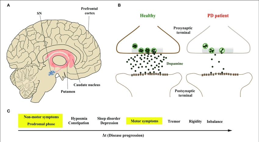
Parkinson's disease (PD), also called idiopathic Parkinson's syndrome or Morbus Parkinson, is a slowly progressive disease of the nervous system. Nerve cells that produce dopamine are gradually lost in a region of the midbrain called the substantia nigra. Dopamine is a messenger substance (neurotransmitter) that helps control movement, so its lack causes the typical movement symptoms.
Clinicians distinguish a tremor-dominant type, an akinetic-rigid type with slowness and stiffness, and mixed forms. When symptoms begin before the age of 50, the disease is called early-onset (young-onset) PD. Diagnosis is clinical, based on a neurological examination; MRI helps to exclude other causes, and dopamine transporter imaging (DaTSCAN) can help where the picture is unclear. Progression is often described with the Hoehn and Yahr stages 1 to 5. To date no cure is known for Parkinson's disease.
Common symptoms include tremor at rest, slowed movement (bradykinesia), muscle stiffness (rigidity), problems with balance and walking, a softer voice, smaller handwriting, loss of smell, constipation and sleep disturbances.
More details can be found below in our scientific section on Parkinson's disease.
What is the ANOVA PD programme?
At ANOVA IRM in Offenbach, Germany, we offer a programme for people with Parkinson's disease, in addition to their neurological treatment. Our aim is to slow progression; whether the nervous system can also repair itself to a meaningful degree is a question research is still working on. The programme combines four components — autologous mesenchymal stem cell secretome (MSEC), TUDCA, senolytic therapy and gut microbiome support. As in our programmes for ALS and MS, several approaches are combined because no single mechanism is proven.
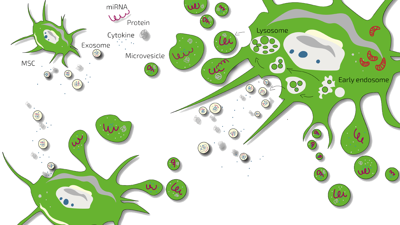
Stem cells are thought to act mainly through soluble factors, together called the secretome, which includes exosomes and many other factors. In Parkinson's disease, the loss of dopamine-producing neurons is accompanied by chronic inflammation in the brain, maintained by the brain's own immune cells (microglia). MSEC is intended to potentially influence this environment and the survival of the remaining nerve cells, not to replace neurons already lost.
ANOVA IRM has not yet tested MSEC in a clinical study. The current state of research is described in the clinical and scientific section below.
The ANOVA PD Programme — Components
MSC Secretome (MSEC)
The signalling molecules released by the patient's own mesenchymal stromal cells, given intrathecally into the cerebrospinal fluid. External laboratory and animal studies suggest they may dampen inflammation and help dopamine-producing neurons survive. Small external clinical studies with MSC products related to MSEC in Parkinson's disease are summarised in the clinical and scientific section below.
TUDCA (tauroursodeoxycholic acid)
A naturally occurring bile acid under investigation for possible neuroprotective effects. In Parkinson's disease the evidence comes from external laboratory and animal studies; why we still use it is set out in the clinical and scientific section below. It is prescribed individually as a pharmacy-compounded preparation (Rezepturarzneimittel) and is not an approved Parkinson's treatment.
Senolytic therapy
Intended to reduce senescent (biologically aged) cells, which release inflammatory signalling molecules. In Parkinson's disease, senescent support cells of the brain (astrocytes) have been described in external laboratory, animal and tissue studies. No senolytic compound is approved for Parkinson's disease, and we do not name individual substances on this website.
Gut microbiome analysis and substitution
Analysis of the gut–brain axis, with personalised dietary or probiotic advice where clinically indicated. External studies report marked differences in the gut microbiome of people with Parkinson's disease, and constipation often begins years before the movement symptoms. That changing the microbiome influences the disease has not been proven by clinical studies.
Which diagnostics do we use for PD at ANOVA IRM?
Which tests are used to diagnose Parkinson's disease?
Parkinson's disease is diagnosed clinically by a neurologist, following the MDS clinical diagnostic criteria. MRI of the brain, with contrast where needed, serves mainly to exclude other causes such as vascular changes or atypical parkinsonian syndromes. Where the diagnosis is uncertain, dopamine transporter imaging (DaTSCAN) and the response to levodopa can help. Severity is tracked with the MDS-UPDRS and the Hoehn and Yahr stages.
What do we review before PD treatment at ANOVA IRM?
Before any treatment decision we review your neurological findings, your disease stage, your current medication and how well it works, and your MRI reports. We also assess whether you are fit for the brief sedation required for tissue collection and, where relevant, add gut microbiome analysis.
- Standard: an MRI of the abdominal wall to exclude a hernia is required before the liposuction, taken no more than four weeks beforehand. It can be done at home or at ANOVA IRM.
- Optional: CT guidance for the intrathecal application at ANOVA IRM, used while the dose is given.
- Optional: further MRI scans of the brain, with or without contrast, where your situation calls for them — for example to follow up a finding on an earlier scan.
Imaging can be carried out in-house; all examinations can also be done by your own doctors. The full diagnostic pathway is described on our diagnostics page.
Why ANOVA IRM for Parkinson's disease?
ANOVA IRM has produced MSEC in its own facility in Offenbach since 2018. Our regulatory status page explains why a tissue preparation made from your own cells, and used only for our own patients, needs no marketing authorisation. Neurological care remains the basis of Parkinson's treatment, and MSEC is offered in addition to it.
What can you expect as a patient with Parkinson's disease?
The first assessment can take place remotely, by phone or video, before any journey to Germany. Treatment itself takes place in person in Offenbach am Main, less than 20 minutes from Frankfurt Airport. Before you decide, you receive an individual written cost estimate.
Quick answers
Is there a cure for Parkinson's disease (PD)?
No. There is currently no cure for Parkinson's disease. Standard care relies on medication such as levodopa, on physiotherapy, occupational and speech therapy and, for suitable patients, on deep brain stimulation.[1] Beyond this, experimental approaches are being explored, among them MSC secretome (MSEC) and the transplantation of dopamine-producing cells grown from stem cells.[22],[23]
Does stem cell therapy cure Parkinson's disease (PD)?
No. Stem cell therapy is not a cure for Parkinson's disease. The use of MSEC at ANOVA IRM aims to slow progression by influencing inflammation in the brain, and it is given alongside — never instead of — your Parkinson's medication.
Is Parkinson's stem cell therapy legal in Germany?
Yes, if the provider holds the official authorisations required for its product. For people with Parkinson's disease, ANOVA IRM produces MSEC under a procurement authorisation (Section 20b AMG) and a manufacturing authorisation under Section 20c AMG; it is administered as an individual treatment attempt. How this fits together is explained on our regulatory status page.
Is Parkinson's stem cell therapy safe?
Not without qualification: every medical treatment carries risks, and your doctor explains them to you in detail before treatment. The quality of the product matters as well. Following the reasoning of the medicines authorities, a secretome made under official authorisation and inspection is to be regarded as safer than one from an unregulated source. Before any treatment, ask the provider which authorisations and certificates it holds. What the authorities warn about is summarised on our page about unproven therapies.
What medication is used for Parkinson's disease (PD)?
Who qualifies, what the process actually involves, and what it costs — step by step.
You are herePD Treatment: Eligibility, Process & Cost
MSEC treatment for Parkinson's disease at ANOVA IRM costs approximately €20,000–€36,000, depending on the number of doses used. It requires a two-day outpatient visit to Offenbach, Germany, for tissue collection, followed by further visits for the applications according to your individual treatment plan. It is available to people with early-to-mid stage Parkinson's disease after individual medical review.

What does the PD treatment timeline look like?
- 01
Initial evaluation
Remote, by phone or video — typically 2 weeks to a few months
- 02
Preliminary screening
In your home country, to avoid an unnecessary journey
- 03
First visit — bloodwork and tissue collection
Two-day outpatient visit to Offenbach: bloodwork first, then — if the results are in order — mini-liposuction under brief sedation
- 04
Production and quality control
Approximately 4 weeks; yields 10 doses per production cycle
- 05
Storage
Up to 2 years, allowing an extended personalised schedule
- 06
Application schedule
Set individually: two doses at the start, then one dose every 4–6 weeks, or two doses every 3 months
Who qualifies for PD treatment, and what are the requirements?
We consider people with a confirmed diagnosis of Parkinson's disease in the early to mid stage whose symptoms persist despite established treatment. Both tremor-dominant and akinetic-rigid forms may be considered. You need to be well enough for mini-liposuction under light sedation, able to breathe independently and comfortably while lying down, and able to travel to Offenbach for repeated applications. Every treatment recommendation follows a full clinical evaluation in which the treating physician weighs benefit and risk, and realistic expectations about the treatment are part of that decision. Applying for an evaluation does not mean that treatment will be offered.
What are the contraindications for PD treatment?
Our treatments are experimental. We treat only people for whom, after medical assessment, we consider the benefit-risk balance to support treatment.
Applies to all ANOVA treatments:
- Active cancer within the last two years
- Under the age of legal majority
- Pregnancy or breastfeeding
- Active infectious disease (hepatitis A, B, C, HIV, syphilis or other)
Specific to Parkinson's disease:
- Unable to breathe unaided, or requiring a ventilator
- Difficulty breathing in the supine position
- Psychiatric disorder that would prevent informed consent or cooperation
What does PD therapy at ANOVA IRM involve, step by step?
What screening is needed before you travel for PD treatment?
Before you travel, a preliminary screening in your home country — usually a blood test for HIV, hepatitis A, B or C, syphilis and other active infections — helps you avoid an unnecessary journey. It also looks for medical factors that would already rule out treatment.
What happens at your first PD visit to Offenbach, Germany?
The first treatment visit takes about two days in Offenbach, on an outpatient basis. On the first day, bloodwork is repeated at ANOVA IRM as German medical rules require, and the physician completes the final assessment. If the results are in order, a small amount of abdominal fat tissue is collected the next day by mini-liposuction under brief sedation. From this point to the first secretome application is usually four to six weeks.
How is your MSEC for PD produced and quality-controlled?
Mesenchymal stromal cells are isolated from the collected tissue and expanded under controlled conditions in our own facility in Offenbach, under official authorisation and officially inspected good professional practice (GFP). GFP is the German counterpart of Good Tissue Practice (GTP) in the US. A registration with the US FDA is, on its own, no official confirmation that the rules are met. In Germany, by contrast, GFP is a condition of the official authorisation and is regularly inspected by the authorities. These cells are then used to produce your MSEC preparation — the signalling molecules they release, not the living cells. Production and quality control take approximately four weeks and yield 10 doses per production cycle. Each patient batch is tested with methods validated according to the European Pharmacopoeia (Ph. Eur.) and GMP, and released under a documented procedure; the released secretome is stored at −80 °C and keeps for up to two years.
How often is MSEC applied in PD?
Because Parkinson's disease is a chronic, progressive disease for which there is currently no cure, we recommend a longer-term treatment using all 10 doses produced rather than a single application; see the timeline above.
The schedule is set individually for each patient, for example:
- At the start: two intrathecal doses.
- If you live close by: a single MSEC dose every 4 to 6 weeks.
- If you live further away: a double dose every 3 months — about five visits over 15 months for the 10 doses produced.
- Standard route for PD: intrathecal, by lumbar puncture into the cerebrospinal fluid. This is intended to bring the secretome closer to the brain than an infusion into the bloodstream, which would have to cross the blood–brain barrier.
- Individual alternative: intravenous, if you specifically wish it or if systemic treatment is medically necessary.
All doses must be used within the two-year shelf life of the stored MSEC; continuing after that requires a new tissue collection and production cycle. Intrathecal applications are standard procedures when performed by experienced physicians; where the anatomy requires it, ANOVA IRM uses CT guidance from its in-house radiology. The benefit-risk assessment of intrathecal administration is set out on a separate page.
What does PD treatment with MSEC cost?
MSEC treatment for Parkinson's disease costs approximately €20,000 to €36,000 and is billed according to the German Medical Fee Schedule (GOÄ). The base package — the mini-liposuction and the first three MSEC doses — costs approximately €20,000; each further dose is charged separately at approximately €2,250 to €2,350 depending on the application route. Because we recommend using all 10 doses over a longer period for a progressive disease such as Parkinson's, most treatment plans reach the upper end of this range. Further examinations, diagnostics and your sedation preferences also affect the total. You receive an individual written estimate before any treatment decision. Our blog post on treatment cost explains what drives the cost of stem cell treatment in general. Travel, accommodation and other personal expenses should be budgeted separately. Health insurance does not ordinarily reimburse this Parkinson's treatment, unless your insurer has explicitly agreed to cover it. All prices are subject to change.
Because Parkinson's disease is chronic and progressive, a double liposuction can be recommended: it yields 20 doses, which can be used over the current shelf life of two years from a single collection. The cost is correspondingly higher and is shown in your individual estimate.
Why does the cost of PD treatment vary with the application route?
All treatments at ANOVA IRM are billed according to the German Medical Fee Schedule (Gebührenordnung für Ärzte, GOÄ); within it, the cost varies mainly with the route of application. For Parkinson's disease we mainly administer MSEC intrathecally, because in our assessment it is most likely to act there: the cerebrospinal fluid is a small, closed compartment that surrounds the brain and spinal cord. We use intravenous administration only if you specifically wish it or if systemic treatment is medically necessary. The exact cost of your treatment plan is set out in your individual written cost estimate.
Frequently asked questions — eligibility, process and travel
How is MSEC administered for Parkinson's disease (PD)?
By intrathecal injection: our physicians in Offenbach give MSEC by lumbar puncture into the cerebrospinal fluid. Two doses are given at the start; after that, one dose every 4 to 6 weeks if you live nearby, or a double dose every 3 months. Intravenous infusion is used only at your specific request or if systemic treatment is medically necessary.
How long does the whole Parkinson's treatment process take?
About one and a half years from first contact to the last dose. The remote assessment takes from about two weeks to several months, depending on appointments and on how quickly your medical records are available. Tissue collection then takes about two days in Offenbach, followed by approximately four weeks of production and quality control. The applications extend over about 15 months and must be completed within the two-year shelf life of the stored MSEC.
Does health insurance cover Parkinson's stem cell therapy in Germany?
No. Health insurers do not ordinarily reimburse experimental MSEC treatment for Parkinson's disease, so you should plan to pay privately. The situation differs only if your insurer has explicitly agreed in advance to cover the programme; please ask your insurer directly.
Should I stop my current PD medication such as levodopa?
No. The programme complements guideline-based care. Please continue to work with your treating neurologist throughout.
Should I continue my existing PD treatment such as levodopa?
Yes. Medication, physiotherapy and your other treatments continue as your neurologist directs; the programme is added to them and changes nothing unless your treating physician recommends a change.
How often do I need to travel to Germany for Parkinson's stem cell treatment?
Usually about five times over 15 months. The first trip, of about two days, is for bloodwork and tissue collection in Offenbach, less than 20 minutes from Frankfurt Airport. The further trips are for the applications — a single dose every 4 to 6 weeks if you live close by, otherwise a double dose every 3 months. The full sequence is described on the MSC secretome page.
What medical records should I send for a Parkinson's assessment?
Please send us your most recent documents; we will ask if we need anything else. Helpful are, for example, your neurologist's letters confirming the diagnosis, your current medication plan with doses and times, and MRI reports and images of the brain. A DaTSCAN report, if one was done, and recent MDS-UPDRS or Hoehn and Yahr assessments also help. Our patient care team will tell you how to send large image files.
Does ANOVA IRM treat international patients with Parkinson's disease?
Yes. People with Parkinson's disease from other countries can begin from home: we advise in English and carry out the first assessment by phone or video.
Parkinson's biology, standard of care, the rationale behind each component and all references.
You are herePD: Clinical & Scientific Evidence
1. What happens in Parkinson's disease (PD)?
Parkinson's disease, first described by James Parkinson in 1817 as the "shaking palsy", is the second most common neurodegenerative disease after Alzheimer's disease.[2] Its hallmark is the loss of dopaminergic neurons in the substantia nigra pars compacta. Inside the affected nerve cells, misfolded alpha-synuclein protein forms deposits called Lewy bodies and Lewy neurites.[2] By the time movement symptoms appear, a large part of these neurons has already been lost.
According to the Braak staging model, Lewy pathology begins in the lower brainstem and in the olfactory system. Over years it ascends to the midbrain and the cortex.[12] This fits the observation that loss of smell (hyposmia), constipation and REM sleep behaviour disorder can precede the movement symptoms by many years.[2] Clinicians distinguish tremor-dominant, akinetic-rigid and mixed types, and early-onset or young-onset PD.[1]
Idiopathic Parkinson's syndrome must be distinguished from atypical parkinsonian syndromes, such as multiple system atrophy (MSA) and progressive supranuclear palsy (PSP), and from drug-induced or vascular parkinsonism.[3] Research into stem cell-based approaches follows two paths. One replaces lost dopamine-producing cells with cells grown from pluripotent stem cells.[21] The other, to which MSEC belongs, investigates signalling molecules that may influence inflammation and the survival of the remaining nerve cells.[14],[15]
What causes Parkinson's disease, and what are the risk factors?
In most people the cause of Parkinson's disease is not known. Researchers assume that ageing, genetic susceptibility and environmental exposures interact; what triggers the disease and what drives its progression are two different questions, and the next section deals with progression.[2]
- Age: the risk rises with age, and Parkinson's disease is uncommon before the age of 50.[1]
- Sex: men are affected about 1.4 to 1.5 times as often as women.[1],[4]
- Genetics: about 3 to 5% of cases have a single genetic cause, for example variants in SNCA, LRRK2 or PRKN; variants in the GBA1 gene raise the risk.[2]
- Environment: exposure to pesticides and head injuries are associated with a higher risk.[2]
What drives the progression of Parkinson's disease?
The cause of Parkinson's disease is unknown, and the drivers of its progression are a separate question. Misfolded alpha-synuclein is thought to spread from cell to cell along nerve pathways, while mitochondrial dysfunction, oxidative stress and impaired protein clearance weaken the affected neurons.[2],[12] Activated microglia and immune cells maintain chronic inflammation in the brain, documented in imaging, blood and cerebrospinal fluid studies.[13] This inflammation is thought to contribute to further loss of neurons, and senescent astrocytes and the gut–brain axis are discussed as further contributors.[29],[30]
This is where our programme aims — at the course of the disease, not at its cause, and not at a cure.
2. What is the guideline-based standard of care for Parkinson's disease (PD)?
Treatment is symptomatic and follows guidelines such as the German S2k guideline on Parkinson's disease.[9] Levodopa, combined with a decarboxylase inhibitor such as carbidopa or benserazide, remains the most effective drug for the motor symptoms. Dopamine agonists such as pramipexole, ropinirole or rotigotine, MAO-B inhibitors such as rasagiline or safinamide, and COMT inhibitors such as entacapone or opicapone are used alone or with levodopa.[1],[9] Amantadine can reduce involuntary movements caused by long-term levodopa treatment (dyskinesias).[1]
When motor fluctuations can no longer be controlled with tablets, device-aided therapies are considered: deep brain stimulation (DBS) of the subthalamic nucleus, levodopa-carbidopa intestinal gel or continuous apomorphine infusion.[1],[9] In an external randomised trial, subthalamic DBS improved quality of life and motor scores more than medication alone, with more serious adverse events.[10] Physiotherapy, occupational therapy and speech and language therapy support mobility, everyday function, speech and swallowing throughout.[1]
None of these treatments has been shown to slow the loss of dopamine-producing neurons or to replace them.[1] That gap is what research into cell-based approaches is exploring; MSEC does not replace standard care.
How is Parkinson's disease diagnosed, and what do we not do ourselves?
The diagnosis of Parkinson's disease is made by neurologists, not by us. It follows the MDS clinical diagnostic criteria: slowed movement (bradykinesia) together with rest tremor, rigidity or both, supported for example by a clear response to dopaminergic medication.[3] MRI of the brain excludes other causes. Dopamine transporter imaging (DaTSCAN, a form of SPECT) helps when parkinsonism is uncertain, and a normal scan argues strongly against Parkinson's disease.[1],[3] An alpha-synuclein seed amplification assay in cerebrospinal fluid is increasingly studied; in a large external cohort study it detected Parkinson's disease with a sensitivity of about 88% and a specificity of about 96%.[11] Severity is recorded with the MDS-UPDRS and the Hoehn and Yahr stages.[5]
What we do ourselves is imaging: MRI of the brain with or without contrast, and CT guidance for an intrathecal application. This is carried out at the Prof. Dr. Stehling Institut für Bildgebende Diagnostik GmbH in the same building, which also belongs to Dr. Stehling. We do not perform DaTSCAN, lumbar puncture for diagnosis or the neurological diagnosis itself; please bring these findings from your neurologist.
3. What is the scientific rationale for PD treatment, component by component?
MSC secretome (MSEC)
ANOVA IRM has not yet tested MSEC in a clinical study. Modern stem cell concepts for Parkinson's disease follow two paths. Cell replacement uses dopamine-producing cells derived from embryonic or induced pluripotent stem cells, transplanted surgically into the brain.[21] MSEC follows the second path: it uses the signalling molecules of mesenchymal stromal cells — growth factors, cytokines and extracellular vesicles — rather than living cells.[14],[15]
In an external systematic review of 13 animal studies, extracellular vesicles from mesenchymal stem cells improved motor tests, lowered inflammatory markers and preserved dopamine-producing neurons.[14] In an external animal study in rats, the secretome of human mesenchymal stem cells, injected into the brain, increased the number of dopaminergic neurons and improved motor performance.[15] These are laboratory and animal data, and they cannot be transferred directly to people.
Small external clinical studies have tested MSC products — cells, not secretome — in Parkinson's disease. In a small external open-label study of bone marrow-derived cells in seven patients, three improved steadily; there was no control group.[16] A small external phase 1 trial with intravenous donor cells in 20 patients reported no serious reactions.[17] A small external randomised phase 2 trial in 45 patients gave mixed results: three infusions showed a numerical advantage over placebo, whereas two infusions did worse than placebo.[18] In a second small external randomised phase 2 trial, with fat-derived cells in 24 patients, a clear treatment effect could not yet be demonstrated.[19]
Small external phase 1 and phase 1/2 trials of transplanted dopamine-producing cells, grown from embryonic or induced pluripotent stem cells, reported no serious cell-related safety problems and signs of graft survival on PET imaging.[22],[23],[24] These are different products from MSEC, which contains no cells. The International Parkinson and Movement Disorder Society considers the evidence insufficient for the widespread use of stem cell therapies outside carefully controlled clinical trials.[20]
What is the efficacy hypothesis behind MSC secretome in PD?
In Parkinson's disease, the dopamine-producing neurons of the substantia nigra die gradually, and activated microglia and astrocytes maintain an inflammatory environment around them.[13] The hypothesis is that the soluble factors of the secretome — among them neurotrophic factors such as BDNF and GDNF — influence this environment, dampen inflammation and support the survival of the remaining neurons.[14],[15]
A possible endpoint for testing this hypothesis would be the rate of change on the motor part of the MDS-UPDRS. One limitation belongs with it: neurons already lost are not expected to be replaced. Our aim is to slow progression, not to cure the disease.
TUDCA
TUDCA is a naturally occurring bile acid studied for effects on mitochondrial function, oxidative stress and programmed cell death. In an external animal study in a mouse model of Parkinson's disease, TUDCA given before the neurotoxin MPTP protected dopaminergic neurons.[27]
No clinical trial of TUDCA in Parkinson's disease has been completed. A small external randomised phase 2 trial tested the related bile acid UDCA in 30 people with Parkinson's disease. It was well tolerated, but an effect on motor scores on the MDS-UPDRS part III could not yet be demonstrated compared with placebo.[28] Whether TUDCA changes the course of Parkinson's disease therefore remains open.
Why do we still use TUDCA? Talk to us about it.
Cellular senescence and senolytics
Senescent cells stop dividing but continue releasing inflammatory signalling molecules. In external laboratory and animal studies, the pesticide paraquat, a suspected environmental risk factor for Parkinson's disease, made astrocytes senescent. Markers of senescent astrocytes were also found in brain tissue from people with Parkinson's disease.[29] In mice, removing senescent cells reduced the damage to dopaminergic neurons caused by paraquat.[29] The evidence is preclinical; no senolytic compound is approved for Parkinson's disease, and we do not name individual substances on this website.
Gut microbiome
The gut–brain axis is a major field of Parkinson's research. Constipation often begins years before the movement symptoms, and the vagus nerve connects the gut with the brainstem region where, according to Braak, Lewy pathology begins.[2],[12] A large external metagenomic study of 490 people with Parkinson's disease and 234 controls found widespread changes in gut bacteria and their functions.[31] In an external animal study, gut bacteria were required for the motor deficits, and bacteria from people with Parkinson's disease worsened them.[30]
Two small external randomised phase 2 trials of faecal microbiota transplantation came to different results. One found a mild but lasting improvement in motor scores after 12 months; in the other, a clinically meaningful benefit could not yet be demonstrated.[32],[33] ANOVA IRM offers microbiome analysis as part of the individual assessment. No probiotic or dietary intervention is established as a treatment for Parkinson's disease.
Talk to us about current changes or additions to the programme. For a number of conditions ANOVA IRM also offers infusion therapies alongside the components described above.
4. Where does our own PD evidence stand?
We have no evaluated dataset of our own for Parkinson's disease yet. What we can say about MSEC in Parkinson's disease therefore rests on the published literature and on the rationale described above. Personal accounts from patients are collected in our testimonials; they are individual reports, not outcome data.
Stem cell-based therapy for Parkinson's disease remains experimental, and outcomes vary from patient to patient. Treatment goals are agreed individually and monitored through clinical and functional measures: motor symptoms such as tremor, rigidity and slowed movement, mobility and everyday function, non-motor symptoms and quality of life. Treatment cannot guarantee slower progression, relief of symptoms or the replacement of lost nerve cells.
What is the realistic goal of PD treatment?
Current Parkinson's therapy aims to control symptoms and to preserve function for as long as possible; no treatment has yet been shown to slow the underlying loss of neurons.[1] For MSEC, too, the aim is to slow progression, not to reverse damage that has already occurred. Research into cell replacement is advancing, but no one can honestly promise a date for a cure.
5. Legal framework for MSEC in PD
MSEC is an autologous tissue preparation made from the patient's own cells. For its production, ANOVA IRM holds a procurement authorisation (Section 20b AMG) and a manufacturing authorisation under Section 20c AMG, both issued and regularly inspected by the Hessian State Office for Health and Care (HLfGP). The individual treatment attempt (individueller Heilversuch) refers only to the administration and follows full disclosure and informed consent. As the product is used only for ANOVA IRM's own patients, it needs no authorisation under Section 4b, no marketing authorisation under Section 21 and no approval under Section 21a AMG. The full derivation is on our regulatory status page.
6. Frequently asked questions — Parkinson's disease (PD), evidence and approval status
Is stem cell therapy approved for Parkinson's disease (PD) anywhere?
Yes, in one country and only conditionally. In Japan, raguneprocel (AMCHEPRY) — dopaminergic progenitor cells grown from donor-derived induced pluripotent stem cells — received conditional, time-limited approval in March 2026; full approval depends on further data.[25] It is transplanted surgically into the brain to replace lost cells, a different approach from ANOVA's cell-free MSEC. In Germany, the EU and the US, no stem cell therapy is approved for Parkinson's disease; a phase 3 trial of a comparable cell product is under way.[26]
Is MSEC the same as dopamine cell transplantation for Parkinson's disease (PD)?
No. MSEC is a cell-free preparation of the signalling molecules released by mesenchymal stromal cells from the patient's own fat tissue. In the trials of cell replacement, dopamine-producing cells grown from embryonic or induced pluripotent stem cells are transplanted surgically into the brain to replace lost neurons.[22],[23],[24] ANOVA IRM does not perform this kind of cell transplantation.
Are MSC studies evidence that MSEC works in Parkinson's disease (PD)?
No. External laboratory, animal and small clinical studies with MSC products provide a rationale and indirect evidence for Parkinson's disease, but no proof. Proof would require a randomised controlled phase 3 trial with MSEC itself, and none exists. Why we use a cell-free product is explained on the MSC secretome page.
What type of stem cell therapy does ANOVA use for Parkinson's disease (PD)?
ANOVA uses an autologous MSC secretome (MSEC), produced from mesenchymal stromal cells taken from the patient's own abdominal fat tissue. The preparation given is cell-free and contains no living cells.
What is the life expectancy with Parkinson's disease (PD)?
Many people live with Parkinson's disease for many years, often decades, but life expectancy is on average reduced.[2] In external cohort studies that followed people from diagnosis, mortality was about 1.5 times that of the general population, and age and dementia were the main predictors of death.[6] Life expectancy is shortened mainly when parkinsonism is diagnosed before the age of 70.[7] The individual outlook depends on age at diagnosis, overall health, the course of the disease and complications affecting mobility or swallowing.
How common is Parkinson's disease (PD)?
About 6.1 million people worldwide had Parkinson's disease in 2016, and a modelling study based on the Global Burden of Disease Study 2021 projects 25.2 million by 2050, mainly because populations are ageing.[2],[4] Parkinson's disease is uncommon before the age of 50, and its prevalence peaks between the ages of 85 and 89.[1] About a quarter of people develop it before the age of 65, and 5 to 10% before 50.[2] Men are affected about 1.4 to 1.5 times as often as women.[1],[4]
What are the symptoms of Parkinson's disease (PD)?
The main symptoms of Parkinson's disease are tremor at rest, slowed movement (bradykinesia), muscle stiffness (rigidity) and problems with balance, posture and walking. Early signs can be subtle and often affect only one side of the body at first. The combination, severity and progression of symptoms vary considerably from person to person. Because every symptom can also have other causes, a neurologist must make the diagnosis.
Movement-related symptoms
- tremor at rest, often beginning in one hand, sometimes as a rolling movement of thumb and index finger ("pill-rolling")
- slowed movement and difficulty with automatic movements, such as a reduced arm swing
- stiff, rigid muscles
- a stooped or forward-leaning posture, balance problems and falls
- smaller handwriting, especially towards the end of words and sentences (micrographia)
- reduced facial expression (hypomimia, "masked face")
- a softer, more monotonous voice and other changes in speech
Non-motor symptoms
- loss of smell (hyposmia) and constipation, often years before the movement symptoms[2]
- sleep disturbances, including acting out dreams with sudden movements (REM sleep behaviour disorder)
- fatigue, depression and anxiety
- dizziness or fainting on standing up (orthostatic hypotension)
- pain, bladder problems and sexual dysfunction
- changes in memory and thinking, and difficulty swallowing (dysphagia) in later stages
Which forms of Parkinson's disease (PD) are there, and how does it progress?
Parkinson's disease is divided by its main symptoms into a tremor-dominant, an akinetic-rigid and a mixed type, and by age at onset into early-onset and later-onset forms.[1] Its progression is commonly described with the five Hoehn and Yahr stages.[5]
- Tremor-dominant type: tremor is the leading symptom; slowness and stiffness are less marked.
- Akinetic-rigid type: slowed movement, stiffness and, later, problems with posture and walking dominate, with little or no tremor.
- Mixed type: tremor, slowness and stiffness are roughly equally pronounced.
- Early-onset (young-onset) PD: symptoms begin before the age of 50; genetic causes are more common in this group.[2]
The Hoehn and Yahr stages:
- Stage 1: mild movement symptoms on one side of the body, with little impact on daily activities.
- Stage 2: symptoms on both sides of the body, with balance generally preserved; daily tasks may take longer.
- Stage 3: balance problems and falls may emerge, although many people can still live independently.
- Stage 4: symptoms are severely limiting, and substantial help is needed for everyday activities.
- Stage 5: standing or walking may no longer be possible without assistance, and continuous care may be required.
This scale mainly describes motor disability. Parkinson's disease does not progress at the same rate or in the same sequence in everyone.
What complications can Parkinson's disease (PD) cause?
Parkinson's disease can lead to falls and fractures, swallowing problems with a risk of choking, dementia, hallucinations and depression. Drops in blood pressure on standing, bladder problems and excessive daytime sleepiness are also common in later years. With long-term levodopa treatment, motor fluctuations and involuntary movements (dyskinesias) can develop.[1] In a long-term external cohort study, 83% of the people still alive 20 years after diagnosis had dementia and 87% had falls; almost half had choking episodes.[8]
Important Pages
Questions people usually ask next. Each is answered in full on its own page.
- Who are we?
- Is ANOVA legal?
- Is stem cell therapy safe?
- What is MSC secretome (MSEC)?
- What is BMC (Bone Marrow Concentrate)?
- What are stem cells?
- What should I ask when looking for a stem cell treatment?
- Which conditions do we treat?
- Benefit-risk assessment of intrathecal administration
- Does the secretome pass the blood–brain barrier?
- Parkinson's disease and MSC secretome — research background
- Parkinson's disease — further treatment approaches
Affiliations of ANOVA
ANOVA IRM shares its premises in Offenbach with two further institutions owned by Dr. Stehling: the Institut für Bildgebende Diagnostik (IBDO), providing MRI and CT imaging, and the Vitus Prostate Center. Because imaging is carried out in the same building, condition-specific diagnostics — including CT-guided procedures for the intrathecal application — can be performed in-house rather than referred elsewhere. See diagnostics at ANOVA IRM.
References — Parkinson's disease-specific literature
Parkinson's disease — overview, diagnosis and standard care
- [1] Armstrong MJ, Okun MS. Diagnosis and treatment of Parkinson disease: a review. JAMA. 2020;323(6):548–560. doi:10.1001/jama.2019.22360. PMID: 32044947.
- [2] Bloem BR, Okun MS, Klein C. Parkinson's disease. Lancet. 2021;397(10291):2284–2303. doi:10.1016/S0140-6736(21)00218-X. PMID: 33848468.
- [3] Postuma RB, Berg D, Stern M, et al. MDS clinical diagnostic criteria for Parkinson's disease. Mov Disord. 2015;30(12):1591–1601. doi:10.1002/mds.26424. PMID: 26474316.
- [4] Su D, Cui Y, He C, et al. Projections for prevalence of Parkinson's disease and its driving factors in 195 countries and territories to 2050: modelling study of Global Burden of Disease Study 2021. BMJ. 2025;388:e080952. doi:10.1136/bmj-2024-080952. PMID: 40044233.
- [5] Hoehn MM, Yahr MD. Parkinsonism: onset, progression and mortality. Neurology. 1967;17(5):427–442. doi:10.1212/wnl.17.5.427. PMID: 6067254.
- [6] Macleod AD, Taylor KS, Counsell CE. Mortality in Parkinson's disease: a systematic review and meta-analysis. Mov Disord. 2014;29(13):1615–1622. doi:10.1002/mds.25898. PMID: 24821648.
- [7] Dommershuijsen LJ, Heshmatollah A, Darweesh SKL, et al. Life expectancy of parkinsonism patients in the general population. Parkinsonism Relat Disord. 2020;77:94–99. doi:10.1016/j.parkreldis.2020.06.018. PMID: 32712564.
- [8] Hely MA, Reid WG, Adena MA, et al. The Sydney multicenter study of Parkinson's disease: the inevitability of dementia at 20 years. Mov Disord. 2008;23(6):837–844. doi:10.1002/mds.21956. PMID: 18307261.
- [9] Höglinger G, Trenkwalder C, et al. Parkinson-Krankheit, S2k-Leitlinie, 2023. In: Deutsche Gesellschaft für Neurologie (Hrsg.), Leitlinien für Diagnostik und Therapie in der Neurologie. AWMF-Registernummer 030-010, Version 8.1. https://register.awmf.org/de/leitlinien/detail/030-010
- [10] Deuschl G, Schade-Brittinger C, Krack P, et al. A randomized trial of deep-brain stimulation for Parkinson's disease. N Engl J Med. 2006;355(9):896–908. doi:10.1056/NEJMoa060281. PMID: 16943402.
- [11] Siderowf A, Concha-Marambio L, Lafontant DE, et al. Assessment of heterogeneity among participants in the Parkinson's Progression Markers Initiative cohort using α-synuclein seed amplification: a cross-sectional study. Lancet Neurol. 2023;22(5):407–417. doi:10.1016/S1474-4422(23)00109-6. PMID: 37059509.
Disease mechanisms
- [12] Braak H, Del Tredici K, Rüb U, et al. Staging of brain pathology related to sporadic Parkinson's disease. Neurobiol Aging. 2003;24(2):197–211. doi:10.1016/S0197-4580(02)00065-9. PMID: 12498954.
- [13] Tansey MG, Wallings RL, Houser MC, et al. Inflammation and immune dysfunction in Parkinson disease. Nat Rev Immunol. 2022;22(11):657–673. doi:10.1038/s41577-022-00684-6. PMID: 35246670.
MSC, secretome and cell-based therapies
- [14] Wang XS, Wang Y, Xu Y, et al. Effectiveness of mesenchymal stem cell-derived extracellular vesicles therapy for Parkinson's disease: a systematic review of preclinical studies. World J Stem Cells. 2025;17(4):102421. doi:10.4252/wjsc.v17.i4.102421. PMID: 40308882.
- [15] Teixeira FG, Carvalho MM, Panchalingam KM, et al. Impact of the secretome of human mesenchymal stem cells on brain structure and animal behavior in a rat model of Parkinson's disease. Stem Cells Transl Med. 2017;6(2):634–646. doi:10.5966/sctm.2016-0071. PMID: 28191785.
- [16] Venkataramana NK, Kumar SKV, Balaraju S, et al. Open-labeled study of unilateral autologous bone-marrow-derived mesenchymal stem cell transplantation in Parkinson's disease. Transl Res. 2010;155(2):62–70. doi:10.1016/j.trsl.2009.07.006. PMID: 20129486.
- [17] Schiess M, Suescun J, Doursout MF, et al. Allogeneic bone marrow-derived mesenchymal stem cell safety in idiopathic Parkinson's disease. Mov Disord. 2021;36(8):1825–1834. doi:10.1002/mds.28582. PMID: 33772873.
- [18] Schiess MC, Suescun J, Martinez-Lemus JD, et al. Allogeneic bone marrow-derived mesenchymal stem cells for Parkinson's disease: a randomized trial. Mov Disord. 2025;40(12):2688–2697. doi:10.1002/mds.70028. PMID: 40891094.
- [19] Vij R, Kim H, Park H, et al. Evaluation of multiple intravenous infusions of autologous adipose-derived mesenchymal stem cells in Parkinson's disease: a randomized, double-blind clinical trial. Parkinsons Dis. 2026;2026:9934417. doi:10.1155/padi/9934417. PMID: 42137372 (ClinicalTrials.gov NCT04928287).
- [20] International Parkinson and Movement Disorder Society, Scientific Issues Committee. MDS Position Paper — Use of Stem Cell Therapies for Parkinson's Disease. January 2021. Accessed 28 September 2026. https://www.movementdisorders.org/MDS/News/News--Notices/News-Release---Stem-Cell-Therapies/MDS-Position-Paper-Use-of-Stem-Cell-Therapies-for-Parkinsons-Disease.htm
- [21] Skidmore S, Barker RA. Challenges in the clinical advancement of cell therapies for Parkinson's disease. Nat Biomed Eng. 2023;7(4):370–386. doi:10.1038/s41551-022-00987-y. PMID: 36635420.
- [22] Tabar V, Sarva H, Lozano AM, et al. Phase I trial of hES cell-derived dopaminergic neurons for Parkinson's disease. Nature. 2025;641:978–983. doi:10.1038/s41586-025-08845-y. PMID: 40240592.
- [23] Sawamoto N, Doi D, Nakanishi E, et al. Phase I/II trial of iPS-cell-derived dopaminergic cells for Parkinson's disease. Nature. 2025;641:971–977. doi:10.1038/s41586-025-08700-0. PMID: 40240591.
- [24] Paul G, Bjartmarz H, Kirkeby A, et al. Human embryonic stem cell-derived dopaminergic cells for Parkinson's disease: a phase 1/2 open-label trial. Nat Med. 2026;32(9):3449–3457. doi:10.1038/s41591-026-04525-0. PMID: 42426223.
Approval status
- [25] Sumitomo Pharma Co., Ltd. Press release: conditional and time-limited approval in Japan of AMCHEPRY (raguneprocel), allogeneic iPS cell-derived dopaminergic progenitor cells, for Parkinson's disease. 6 March 2026. Accessed 28 September 2026. https://www.sumitomo-pharma.com/news/20260306.html
- [26] BlueRock Therapeutics. exPDite-2: a phase 3 study of bemdaneprocel in Parkinson's disease. ClinicalTrials.gov NCT06944522. Accessed 28 September 2026. https://clinicaltrials.gov/study/NCT06944522
TUDCA and bile acids
- [27] Castro-Caldas M, Carvalho AN, Rodrigues E, et al. Tauroursodeoxycholic acid prevents MPTP-induced dopaminergic cell death in a mouse model of Parkinson's disease. Mol Neurobiol. 2012;46(2):475–486. doi:10.1007/s12035-012-8295-4. PMID: 22773138.
- [28] Payne T, Appleby M, Buckley E, et al. A double-blind, randomized, placebo-controlled trial of ursodeoxycholic acid (UDCA) in Parkinson's disease. Mov Disord. 2023;38(8):1493–1502. doi:10.1002/mds.29450. PMID: 37246815.
Cellular senescence
- [29] Chinta SJ, Woods G, Demaria M, et al. Cellular senescence is induced by the environmental neurotoxin paraquat and contributes to neuropathology linked to Parkinson's disease. Cell Rep. 2018;22(4):930–940. doi:10.1016/j.celrep.2017.12.092. PMID: 29386135.
Gut microbiome
- [30] Sampson TR, Debelius JW, Thron T, et al. Gut microbiota regulate motor deficits and neuroinflammation in a model of Parkinson's disease. Cell. 2016;167(6):1469–1480.e12. doi:10.1016/j.cell.2016.11.018. PMID: 27912057.
- [31] Wallen ZD, Demirkan A, Twa G, et al. Metagenomics of Parkinson's disease implicates the gut microbiome in multiple disease mechanisms. Nat Commun. 2022;13(1):6958. doi:10.1038/s41467-022-34667-x. PMID: 36376318.
- [32] Bruggeman A, Vandendriessche C, Hamerlinck H, et al. Safety and efficacy of faecal microbiota transplantation in patients with mild to moderate Parkinson's disease (GUT-PARFECT): a double-blind, placebo-controlled, randomised, phase 2 trial. EClinicalMedicine. 2024;71:102563. doi:10.1016/j.eclinm.2024.102563. ClinicalTrials.gov NCT03808389.
- [33] Scheperjans F, Levo R, Bosch B, et al. Fecal microbiota transplantation for treatment of Parkinson disease: a randomized clinical trial. JAMA Neurol. 2024;81(9):925–938. doi:10.1001/jamaneurol.2024.2305. PMID: 39073834.
General MSC, extracellular vesicle and secretome literature is maintained on the MSC secretome page; bone marrow literature on the BMC page.