

Research News
Sjögren’s Disease & Dry Mouth:
The Immune Attack and How We Aim to Regain Gland Function
Persistent dry mouth (xerostomia) and dry eyes are the hallmark of Sjögren’s. Here we explain what happens inside the salivary glands, how the damage is measured, which treatments exist today, and how cell-based medicine intends to help the remaining gland tissue regain function.
In short: Sjögren’s disease is a chronic autoimmune disease in which immune cells attack the saliva- and tear-producing glands. It affects about 6 in 10,000 people, about ten women for every man.[1] Today’s standard therapy relieves dryness and controls inflammation; it does not rebuild lost gland tissue. Cell-based approaches that calm the immune attack and support the gland’s own stem cells are under investigation and remain experimental.
Patient and physician information · Read time: about 10 minutes
| Component | Detail |
|---|---|
| Two products | Bone Marrow Concentrate (BMC) and Mesenchymal Stem Cell Secretome (MSEC), each manufactured under official authorisation; no marketing authorisation (experimental) |
| Authorisations | BMC § 20b / § 13 AMG · MSEC § 20b / § 20c AMG (German Medicines Act) |
| Location | Offenbach am Main, Germany |
Plain facts
- Experimental: cell-based treatment of Sjögren’s is not an approved standard therapy in Germany or the EU.
- Not a cure: glands replaced by scar tissue and fat cannot yet be restored.
- Alongside standard care: complements, never replaces, care by your rheumatologist.
- Individually assessed: every recommendation follows diagnostics and informed consent.
The disease, briefly
Sjögren’s is the second most common systemic rheumatic autoimmune disease after rheumatoid arthritis. It mainly attacks the salivary and tear glands, causing dry mouth and dry eyes, but can involve joints, nerves, lungs and kidneys.[2]
| Figure | What it means |
|---|---|
| About 6 in 10,000 people | Pooled prevalence of primary Sjögren’s is about 61 per 100,000; population-based studies give about 43 per 100,000.[1] Single studies report between 22 and 770 per 100,000, mostly because of differences in study design and classification criteria.[3] |
| About 10 : 1 women to men | The mean age of patients is about 56.[1] Symptoms usually begin between the ages of 34 and 57; new cases are most frequent from the age of 65.[3] |
| Under-diagnosed | Because classification criteria are applied inconsistently, Sjögren’s is probably under-diagnosed, possibly most of all in men.[3] |
What causes it
The exact cause is unknown. Sjögren’s develops in several steps: a genetically susceptible person meets an environmental trigger, against a hormonal background that favours women.[4]
- Genetics: HLA genes and interferon and B-cell genes such as IRF5, STAT4, BLK and CXCR5[5]
- Viral triggers: viral infections are thought to activate innate immune sensors in the gland[4],[6]
- Hormones: hormonal factors help explain why women are affected far more often[4]
- Interferon signature: found in many patients, linking alarm signals to B-cell activation[4],[6]
Dry mouth: more than a nuisance
Saliva protects teeth, gums and the lining of the mouth and makes chewing, swallowing and speaking possible. Chronic dry mouth leads to caries, oral thrush, burning tongue and difficulty eating. Not every dry mouth is Sjögren’s: medication, radiation therapy and age are common causes too, which is why proper diagnostics matter. Causes, standard care and our treatment programme are described on our dry mouth page.
Primary Sjögren’s occurs alone; secondary Sjögren’s accompanies rheumatoid arthritis, lupus or systemic sclerosis.[2]
What happens inside the salivary gland
The gland’s own epithelium is both the target and an active initiator of the attack: stressed gland cells call in immune cells, which then destroy the secretory tissue.[7]
The attack, step by step
Most of the glandular epithelium consists of saliva-producing acinar cells; ductal cells transport and modify the saliva. The following steps are described in current reviews.[7],[6]
- 01
Epithelial alarm — gland epithelial cells
Cells sense viral or endogenous RNA/DNA via Toll-like receptors, die by apoptosis and expose the autoantigens Ro/SSA and La/SSB.
- 02
Interferon surge — plasmacytoid dendritic cells
Immune complexes of these antigens trigger a large release of interferon-α.
- 03
T cells arrive — CD4⁺ T helper cells
Gland cells present antigen and release chemokines; Th1, Th17 and T follicular helper cells gather around the ducts.
- 04
B cells organise — B cells, plasma cells
CXCL13 and BAFF draw in B cells, which form ectopic germinal centres and produce autoantibodies.
- 05
Gland cells are killed — CD8⁺ cytotoxic T cells
Killer T cells destroy acinar and ductal cells via perforin, granzyme and Fas ligand.
Who attacks what
| Immune cell | Target / action |
|---|---|
| Plasmacytoid dendritic cells | IFN-α keeps the gland in a permanent “antiviral” alarm state |
| Dendritic cells, macrophages | Present autoantigens; release TNF-α, IL-1, IL-6 |
| CD4⁺ Th1 / Th17 cells | IFN-γ and IL-17 damage acinar cells and tight junctions |
| T follicular helper cells | IL-21 and CD40L drive B-cell maturation |
| CD8⁺ cytotoxic T cells | Directly kill acinar and ductal cells |
| B cells / plasma cells | Anti-Ro/SSA, anti-La/SSB, rheumatoid factor, anti-M3 receptor antibodies |
| NK cells | IFN-γ and cytotoxicity toward stressed epithelium |
How the gland deteriorates
Saliva loss starts as a functional block and becomes structural damage over years. Early on the gland secretes poorly although many acini survive; later the acini are lost and replaced by scar tissue and fat.[7]
- 01
Functional block
Cytokines and anti-M3 antibodies blunt the nerve signal; the water channel aquaporin-5 is misplaced.
- 02
Lymphocyte foci
Clusters of 50 or more lymphocytes form around the ducts and grow in number and size.
- 03
Duct lesions
B cells infiltrate the ducts, forming lymphoepithelial lesions typical of advanced disease.
- 04
Acinar loss
Acini die faster than they are replaced; the gland’s progenitor cells become senescent.
- 05
Fibrosis and fat
Collagen and fat fill the space. Largely irreversible, which is why late dryness responds poorly to drugs.
Watch for lymphoma risk: chronic B-cell stimulation raises the risk of non-Hodgkin lymphoma several times above that of the general population. Persistent gland swelling, low C4 and cryoglobulins are warning signs.[2],[8]
How it is measured
Damage is measured on four levels: secretion, tissue, imaging and blood. Secretion, tissue and antibodies form the 2016 ACR/EULAR classification score; Sjögren’s is classified with 4 points or more.[9]
2016 ACR/EULAR criteria
| Item | Threshold | Points |
|---|---|---|
| Minor salivary gland biopsy | Focus score ≥ 1 per 4 mm² | 3 |
| Anti-SSA/Ro antibodies | Positive | 3 |
| Ocular staining score | ≥ 5 in at least one eye | 1 |
| Schirmer test | ≤ 5 mm in 5 minutes | 1 |
| Unstimulated saliva flow | ≤ 0.1 mL/min | 1 |
What each test shows
- Saliva flow measures how much the glands still secrete.[9]
- Biopsy focus score counts lymphocyte clusters of 50 or more cells; atrophy, fibrosis, fat and germinal centres signal advanced disease.[7],[9]
- Gland ultrasound (OMERACT 0–3): a score of 2 or more in at least one gland gave 72% sensitivity and 91% specificity. Non-invasive and well suited to follow-up.[10]
- Blood: anti-Ro/La, rheumatoid factor, ANA, IgG, C3/C4, cryoglobulins, interferon signature.
- Activity scores: ESSDAI (12 organ domains, main trial endpoint) and ESSPRI (patient-rated dryness, fatigue, pain).[11]
Diagnostics under one roof
ANOVA IRM shares its building in Offenbach with the Institut für Bildgebende Diagnostik (IBDO), so MRI, CT and ultrasound are performed in-house rather than referred elsewhere. See diagnostics at ANOVA IRM.
Standard medications and treatments
Treatment today is mostly symptomatic for dryness, with immunosuppression reserved for organ involvement.[11] In the EU and US no drug is yet approved to modify the disease itself.
| Target | Treatment | Notes |
|---|---|---|
| Dry mouth, mild | Sugar-free gum and lozenges, saliva substitutes, fluoride, dental hygiene | First line; prevents caries and candida |
| Dry mouth, moderate | Pilocarpine or cevimeline (muscarinic agonists) | Work only while acini remain; sweating, flushing |
| Dry eyes | Artificial tears, topical ciclosporin, short topical steroids, autologous serum drops, punctal plugs | With ophthalmology |
| Fatigue, joint pain, mild systemic | Hydroxychloroquine, analgesics, exercise | Modest effect on dryness |
| Active systemic disease | Glucocorticoids at the lowest dose and shortest time | Bridging therapy |
| Steroid-sparing | Methotrexate, leflunomide, azathioprine, mycophenolate | Chosen by organ involved |
| Severe organ disease | Cyclophosphamide; biologics such as rituximab (off-label), abatacept, belimumab | Specialist centres |
Treatment options according to the EULAR recommendations.[11]
New in 2026
Telitacicept, which blocks the B-cell survival signals BAFF and APRIL, received the first approval specifically for Sjögren’s, in China, in June 2026.[12] In the US, ianalumab received FDA Breakthrough Therapy designation in January 2026, and in September 2026 the CD40L blocker dazodalibep met the primary endpoint of a phase 3 trial.[13],[14],[15] Neither is approved in Europe yet.
Immunosuppression: anti-CD20 antibodies
Anti-CD20 antibodies remove B cells, the cells that drive autoantibodies and germinal centres in the gland. Rituximab missed the primary endpoint in two large randomised trials and is used only off-label for severe disease.[11],[16],[17]
How it works
CD20 sits on B cells from the pre-B to the memory stage, but not on most antibody-producing plasma cells. Rituximab, usually 2 × 1,000 mg IV two weeks apart, removes blood B cells for 6–9 months; gland biopsies show fewer B cells and smaller foci.
Because long-lived plasma cells lack CD20, autoantibody levels barely change. The rise in BAFF after depletion may also favour autoreactive B cells when they return.
Next generation: ianalumab targets the BAFF receptor, removing B cells and blocking their survival signal. In two phase 3 trials with a total of 779 patients, monthly injections significantly reduced disease activity, though the absolute difference from placebo was modest.[18]
What the trials showed
- TEARS (France, 120 patients) missed its primary endpoint[16]
- TRACTISS (UK, 133 patients) missed it too, and was judged not cost-effective[17]
- Glands with advanced damage responded worst
Where it still helps
- Cryoglobulinaemic vasculitis
- Severe parotid swelling, neuropathy
- Sjögren’s-associated lymphoma
An option in the EULAR recommendations for severe systemic disease.[11]
Cellular medicine: aiming to regain gland function
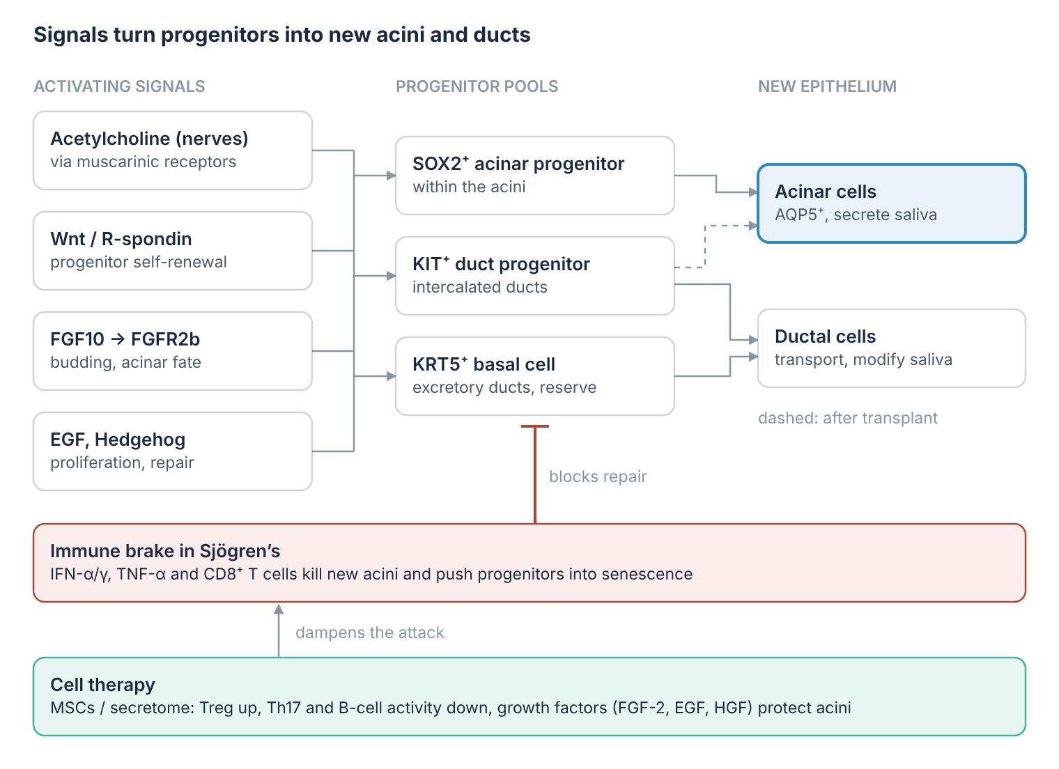
Research suggests that regaining gland function needs two things at once: new secretory epithelium from the gland’s stem and progenitor cells, and a calmer immune environment so the new cells are not destroyed again.[19]
The gland’s own stem cells
| Progenitor | Location | Gives rise to |
|---|---|---|
| SOX2⁺ | Within the acini | New acinar cells |
| KIT⁺ (CD117) | Intercalated ducts | Ducts; after transplant also acini |
| KRT5⁺/KRT14⁺ | Excretory ducts | Duct epithelium, reserve |
| SOX9⁺, ASCL3⁺ | Ducts | Repair after injury |
| Mature acinar cells | Acini | Self-duplication |
Overview of the progenitor pools described in the literature.[19],[20] In Sjögren’s these pools are damaged too: chronic inflammation pushes progenitors into senescence.[21]
How they are activated to form epithelium
- 01
Nerve signal — acetylcholine → SOX2⁺ cells
Parasympathetic nerves keep the progenitor pool alive and drive new acinar cells.
- 02
Wnt / R-spondin — self-renewal
Essential for salivary stem cells to renew themselves.
- 03
FGF10, EGF, Hedgehog — budding and growth
Drive budding, proliferation and acinar fate, as in the embryo.
- 04
Mature epithelium — acinar and ductal cells
Polarised acinar cells with aquaporin-5 and secretory granules, linked to a duct network.
The nerve signal to SOX2⁺ cells was shown in mice after radiation injury; the other signals are described in reviews of salivary gland development and repair.[19],[20]
Cell therapies under investigation
- Gland stem-cell transplant: in irradiated mouse glands, an injection of only 300 to 1,000 KIT⁺ cells improved saliva secretion in 9 of 13 animals.[22] Human studies so far target radiation damage, not Sjögren’s.[19]
- Mesenchymal stromal cells: act mainly through signals; in mice and in an uncontrolled study in patients, donor MSCs given into a vein were followed by more regulatory T cells, fewer Th17 cells and more saliva.[23]
- Direct gland injection: in a randomised, placebo-controlled trial, 74 patients received donor fat-derived MSCs or saline into the parotid glands under ultrasound guidance. Saliva and tear flow were better than with placebo after three months; after six months, saliva flow no longer differed from placebo.[24]
- Cell-free secretome: exosomes and other extracellular vesicles carry the signalling cargo of MSCs, such as microRNAs and growth factors, without living cells. In a mouse model of Sjögren’s, such vesicles dampened the inflammation of the salivary glands.[25]
At ANOVA IRM
For dry mouth, including dry mouth in Sjögren’s, our standard treatment is MSC secretome (MSEC): a cell-free preparation made from the patient’s own fat tissue and injected into the salivary glands under ultrasound guidance. It is manufactured under German regulatory authorisation and official inspection since 2018. Bone marrow concentrate (BMC), our second product, is not used for dry mouth as standard. With MSEC we intend to help the remaining gland tissue regain function; gland tissue that has been replaced by scar and fat is not expected to be restored. Whether MSEC is suitable therefore depends on how much functional gland tissue remains, which is why diagnostics come first. Details are on our dry mouth page.
Senolytics: clearing senescent cells
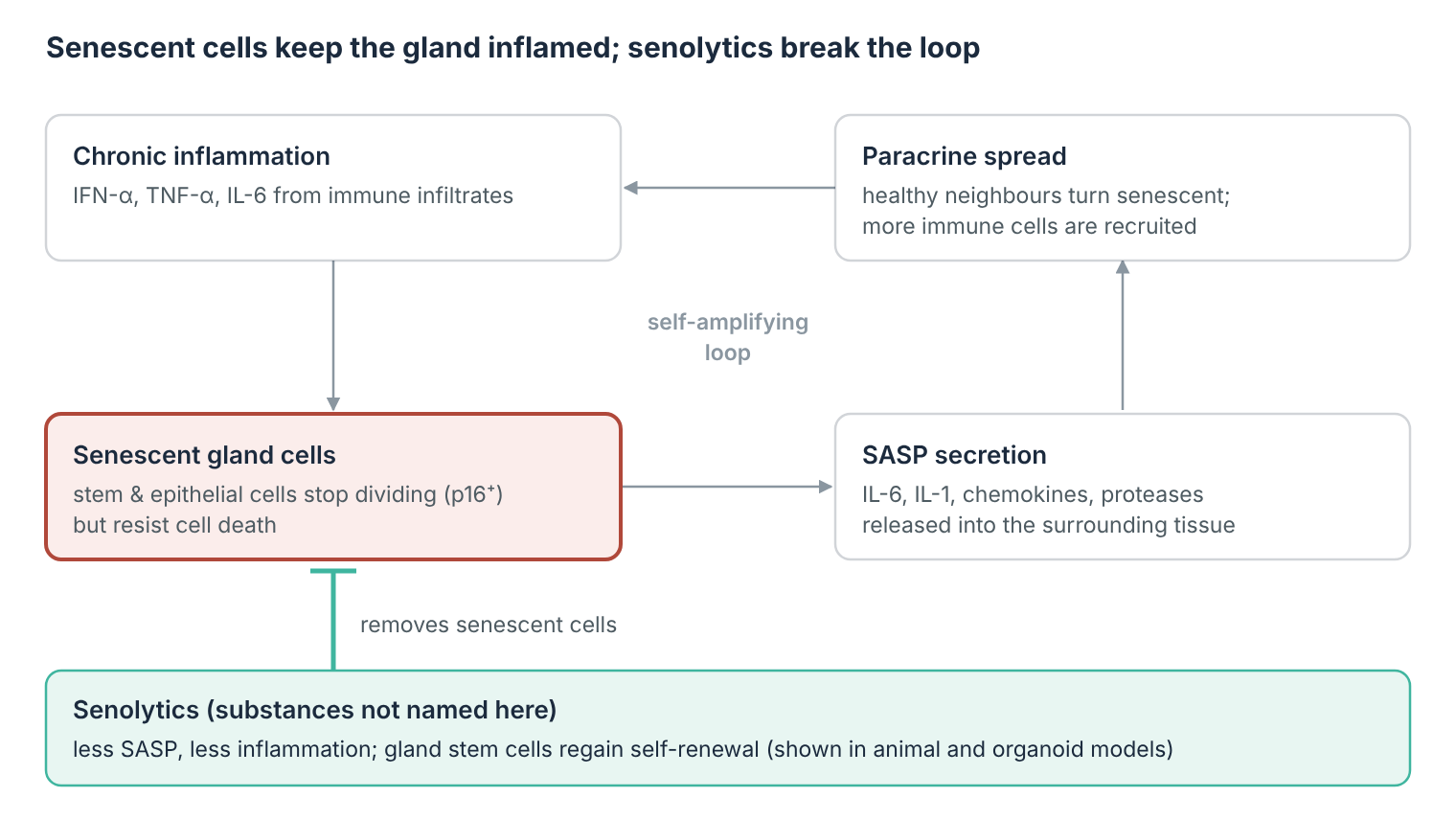
Senescent cells have stopped dividing but refuse to die. They release a cocktail of inflammatory signals that keeps the gland inflamed and pushes healthy neighbouring cells into senescence too. Senolytics are drugs that selectively remove these cells.[26]
Why senescence matters in Sjögren’s
In Sjögren’s, the gland’s own stem cells age prematurely. In biopsies, senescent (p16⁺) cells cluster in the intercalated ducts, exactly where the stem cells live. Patients’ gland stem cells formed about 10 times fewer spheres and up to 5 times fewer organoids than those of healthy donors, and their shortest telomeres were markedly shorter (3.1 vs 5.6 kb), although the patients were younger. A combination of the inflammatory cytokines of Sjögren’s (IFN-α, TNF, IL-6) pushed healthy gland stem cells towards senescence in the lab.[21]
Senescent cells secrete the senescence-associated secretory phenotype (SASP): IL-6, IL-1, chemokines and tissue-degrading proteases. The SASP fuels inflammation and transmits senescence to healthy neighbouring cells, a process called paracrine senescence.[27] In the salivary glands of aged mice, senescence-associated T cells accumulate, attracted by the chemokine CXCL13; in a mouse model of Sjögren’s they accumulate as well, probably via CXCL12.[28]
What senolytics have shown
- Gland stem cells recover. After irradiation, removing senescent cells, genetically or with a senolytic drug, increased the self-renewal of salivary gland stem cells in organoids.[29]
- Saliva is preserved. In mice with radiation-damaged salivary glands, a senolytic drug reduced tissue degeneration and preserved saliva production.[29]
All of this is preclinical. To our knowledge, no senolytic has yet been tested in a clinical trial in Sjögren’s.
How senolytics are used
We do not name individual substances here. Senolytics switch off the survival pathways that keep senescent cells alive; some of them can lower platelet counts. A complementary, non-lytic strategy, called senomorphics, does not kill senescent cells but dampens the SASP.
Because senescent cells accumulate slowly, senolytics are given as brief “hit-and-run” courses rather than continuously. In humans, only nine senolytic trials have been published so far, just two with a control group; no significant safety concerns have emerged, but efficacy is not yet proven.[30]
At ANOVA IRM
Senolytic therapy is already a component of ANOVA’s individualised programme for neurodegenerative disease. In Sjögren’s it would be an experimental, individual therapeutic attempt, considered alongside immunomodulation and cell-based approaches, after full diagnostics and informed consent.
Quick answers
The questions patients with Sjögren’s ask first.
Can stem cell therapy cure Sjögren’s disease?
No. No cell-based treatment has been shown to cure Sjögren’s or to restore gland tissue already replaced by scar and fat. Approaches are investigated for their potential to calm the immune attack and help the remaining gland tissue regain function.
Is cell therapy an approved treatment for Sjögren’s?
No. It is not an approved standard treatment in Germany or the EU. Where offered, it follows individual medical assessment, full disclosure of its experimental nature, and informed consent.
Is every dry mouth Sjögren’s disease?
No. Dry mouth is often caused by medication, radiation therapy for head and neck cancer, or age. Sjögren’s is confirmed by antibodies, a gland biopsy or ultrasound, and saliva and tear tests. If your dry mouth persists for more than three months, it is worth having it checked. More on the causes is on our dry mouth page.
What are senolytics, and are they used in Sjögren’s?
Experimental. Senolytics are drugs that remove senescent cells, which drive inflammation and spread senescence to healthy tissue. In organoid and animal models of radiation damage they improved gland stem-cell function and preserved saliva. They have not yet been tested in clinical trials in Sjögren’s.
Should I stop my current Sjögren’s medication?
No. Any approach at ANOVA complements, not replaces, your current care. Please keep working with your rheumatologist and ophthalmologist.
Can I start with an online appointment?
Yes. The first evaluation takes place by phone or video, without travel. Please have recent findings ready: biopsy, ultrasound, blood tests and current medication. Call +49 (0) 69 50 50 00 944.
Call us for an online appointment
Discuss your diagnosis, findings and options with Dr. mult. Stehling by video, from wherever you are. Call us Monday to Friday on +49 (0) 69 50 50 00 944. Our patient care team arranges the time and tells you which documents to prepare.
- Video consultation in German or English
- Review of your existing findings
- MRI, CT and ultrasound in the same building
- Less than 20 minutes from Frankfurt Airport
Your first contact with our Patient Care Management is free of charge. A consultation with one of our physicians is a paid service; the fee is confirmed before any appointment is booked.
Important Pages
Questions people usually ask next. Each is answered in full on its own page.
- Who are we?
- Is ANOVA legal?
- Is stem cell therapy safe?
- What is MSC secretome (MSEC)?
- What is BMC (Bone Marrow Concentrate)?
- What are stem cells?
- What should I ask when looking for a stem cell treatment?
- Which conditions do we treat?
- Dry mouth (xerostomia) — treatment programme
- Autoimmune diseases — overview
Affiliations of ANOVA
ANOVA IRM shares its premises in Offenbach with two further institutions owned by Dr. Stehling: the Institut für Bildgebende Diagnostik (IBDO), providing MRI and CT imaging, and the VITUS PRIVATKLINIK Prof. Dr. Stehling GmbH. Because imaging is carried out in the same building, condition-specific diagnostics — including ultrasound and MRI of the salivary glands — can be performed in-house rather than referred elsewhere. See diagnostics at ANOVA IRM.
References
- [1] Qin B, Wang J, Yang Z, et al. Epidemiology of primary Sjögren’s syndrome: a systematic review and meta-analysis. Ann Rheum Dis. 2015;74(11):1983–1989. doi:10.1136/annrheumdis-2014-205375. PMID: 24938285.
- [2] Brito-Zerón P, Baldini C, Bootsma H, et al. Sjögren syndrome. Nat Rev Dis Primers. 2016;2:16047. doi:10.1038/nrdp.2016.47. PMID: 27383445.
- [3] Thurtle E, Grosjean A, Steenackers M, et al. Epidemiology of Sjögren’s: a systematic literature review. Rheumatol Ther. 2024;11(1):1–17. doi:10.1007/s40744-023-00611-8.
- [4] Baldini C, Chatzis LG, Fulvio G, et al. Pathogenesis of Sjögren’s disease: one year in review 2024. Clin Exp Rheumatol. 2024;42(12):2336–2343. doi:10.55563/clinexprheumatol/i8iszc. PMID: 39656593.
- [5] Lessard CJ, Li H, Adrianto I, et al. Variants at multiple loci implicated in both innate and adaptive immune responses are associated with Sjögren’s syndrome. Nat Genet. 2013;45(11):1284–1292. doi:10.1038/ng.2792.
- [6] Qi W, Tian J, Wang G, et al. Advances in cellular and molecular pathways of salivary gland damage in Sjögren’s syndrome. Front Immunol. 2024;15:1405126. doi:10.3389/fimmu.2024.1405126.
- [7] Verstappen GM, Pringle S, Bootsma H, Kroese FGM. Epithelial–immune cell interplay in primary Sjögren syndrome salivary gland pathogenesis. Nat Rev Rheumatol. 2021;17(6):333–348. doi:10.1038/s41584-021-00605-2.
- [8] Ansari N, Salesi M. The association between primary Sjogren’s syndrome and non-Hodgkin’s lymphoma: a systematic review and meta-analysis of cohort studies. Clin Rheumatol. 2024;43(7):2177–2186. doi:10.1007/s10067-024-06993-6. PMID: 38722505.
- [9] Shiboski CH, Shiboski SC, Seror R, et al. 2016 American College of Rheumatology/European League Against Rheumatism classification criteria for primary Sjögren’s syndrome: a consensus and data-driven methodology involving three international patient cohorts. Arthritis Rheumatol. 2017;69(1):35–45. doi:10.1002/art.39859. PMID: 27785888.
- [10] Fana V, Dohn UM, Krabbe S, Terslev L. Application of the OMERACT grey-scale ultrasound scoring system for salivary glands in a single-centre cohort of patients with suspected Sjögren’s syndrome. RMD Open. 2021;7(2):e001516. doi:10.1136/rmdopen-2020-001516.
- [11] Ramos-Casals M, Brito-Zerón P, Bombardieri S, et al. EULAR recommendations for the management of Sjögren’s syndrome with topical and systemic therapies. Ann Rheum Dis. 2020;79(1):3–18. doi:10.1136/annrheumdis-2019-216114. PMID: 31672775.
- [12] Vor Bio, RemeGen. Telitacicept receives NMPA approval for the treatment of Sjögren’s disease in China. Press release, 8 June 2026. https://www.biospace.com/press-releases/telitacicept-receives-nmpa-approval-for-the-treatment-of-sjogrens-disease-in-china
- [13] Novartis. Novartis ianalumab receives FDA Breakthrough Therapy designation for Sjögren’s disease. Media release, 16 January 2026. https://www.novartis.com/news/media-releases/novartis-ianalumab-receives-fda-breakthrough-therapy-designation-sjogrens-disease
- [14] Clinical Trials Arena. First of Amgen’s Phase III Sjögren’s disease trials succeeds (OASIZ 301, NCT06104124). 23 September 2026. https://www.clinicaltrialsarena.com/news/first-of-amgens-phase-iii-sjogrens-disease-trials-succeeds/
- [15] American Journal of Managed Care. Pipeline advances in Sjögren disease bring targeted therapies closer. 2 October 2026. https://www.ajmc.com/view/pipeline-advances-in-sj-gren-disease-bring-targeted-therapies-closer
- [16] Devauchelle-Pensec V, Mariette X, Jousse-Joulin S, et al. Treatment of primary Sjögren syndrome with rituximab: a randomized trial (TEARS). Ann Intern Med. 2014;160(4):233–242. doi:10.7326/M13-1085.
- [17] Bowman SJ, Everett CC, O’Dwyer JL, et al. Randomized controlled trial of rituximab and cost-effectiveness analysis in treating fatigue and oral dryness in primary Sjögren’s syndrome (TRACTISS). Arthritis Rheumatol. 2017;69(7):1440–1450. doi:10.1002/art.40093.
- [18] Ianalumab demonstrates significant reduction in disease activity in patients with Sjögren’s disease: efficacy and safety results from two global phase 3 randomized, placebo-controlled, double-blind studies (NEPTUNUS-1 and NEPTUNUS-2). ACR Convergence 2025, abstract. acrabstracts.org
- [19] Salivary gland stem/progenitor cells: advancing from basic science to clinical applications. Review, 2025. PMID: 39856475. PMCID: PMC11759724.
- [20] Emmerson E, May AJ, Berthoin L, et al. Salivary glands regenerate after radiation injury through SOX2-mediated secretory cell replacement. EMBO Mol Med. 2018;10(3):e8051. doi:10.15252/emmm.201708051.
- [21] Pringle S, Wang X, Verstappen GMPJ, et al. Salivary gland stem cells age prematurely in primary Sjögren’s syndrome. Arthritis Rheumatol. 2019;71(1):133–142. doi:10.1002/art.40659. PMID: 29984480.
- [22] Lombaert IMA, Brunsting JF, Wierenga PK, et al. Rescue of salivary gland function after stem cell transplantation in irradiated glands. PLoS One. 2008;3(4):e2063. doi:10.1371/journal.pone.0002063.
- [23] Xu J, Wang D, Liu D, et al. Allogeneic mesenchymal stem cell treatment alleviates experimental and clinical Sjögren syndrome. Blood. 2012;120(15):3142–3151. doi:10.1182/blood-2011-11-391144. PMID: 22927248.
- [24] Li F, Lu J, Shi X, et al. Effect of adipose tissue-derived stem cells therapy on clinical response in patients with primary Sjogren’s syndrome. Sci Rep. 2023;13:13521. doi:10.1038/s41598-023-40802-5.
- [25] Hai B, Shigemoto-Kuroda T, Zhao Q, et al. Inhibitory effects of iPSC-MSCs and their extracellular vesicles on the onset of sialadenitis in a mouse model of Sjögren’s syndrome. Stem Cells Int. 2018;2018:2092315. doi:10.1155/2018/2092315. PMID: 29736173.
- [26] Tang Y, et al. Salivary gland cell senescence in Sjögren’s syndrome: current research and novel therapeutic directions. Immun Ageing. 2025. doi:10.1186/s12979-025-00541-9.
- [27] Acosta JC, Banito A, Wuestefeld T, et al. A complex secretory program orchestrated by the inflammasome controls paracrine senescence. Nat Cell Biol. 2013;15(8):978–990. doi:10.1038/ncb2784.
- [28] Kurosawa M, Shikama Y, Furukawa M, et al. Chemokines up-regulated in epithelial cells control senescence-associated T cell accumulation in salivary glands of aged and Sjögren’s syndrome model mice. Int J Mol Sci. 2021;22(5):2302. doi:10.3390/ijms22052302.
- [29] Peng X, Wu Y, Brouwer U, et al. Cellular senescence contributes to radiation-induced hyposalivation by affecting the stem/progenitor cell niche. Cell Death Dis. 2020;11:854. doi:10.1038/s41419-020-03074-9. PMID: 33056980.
- [30] Khosla S. Translating senolytics from mice to humans. Innov Aging. 2025;9(Suppl 2):igaf122.679. doi:10.1093/geroni/igaf122.679.
This page summarises published research for patient information and does not replace an individual medical consultation.